
- 阻害剤
- 研究分野別
- PI3K/Akt/mTOR
- Epigenetics
- Methylation
- Immunology & Inflammation
- Protein Tyrosine Kinase
- Angiogenesis
- Apoptosis
- Autophagy
- ER stress & UPR
- JAK/STAT
- MAPK
- Cytoskeletal Signaling
- Cell Cycle
- TGF-beta/Smad
- 化合物ライブラリー
- 抗体
- 新製品
- お問い合わせ
CUDC-101
CUDC-101 is a potent multi-targeted inhibitor against HDAC, EGFR and HER2 with IC50 of 4.4 nM, 2.4 nM, and 15.7 nM, and inhibits class I/II HDACs, but not class III, Sir-type HDACs. Phase 1.
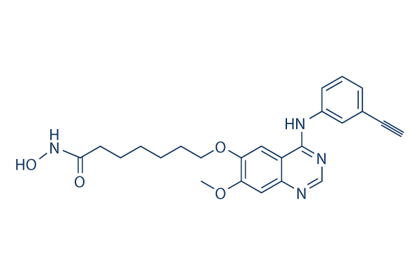
CAS No. 1012054-59-9
文献中Selleckの製品使用例(22)
カスタマーフィードバック3个实验数据
製品安全説明書
現在のバッチを見る:
純度:
99.58%
99.58
CUDC-101関連製品
| 関連ターゲット | HDAC1 HDAC2 HDAC3 HDAC4 HDAC5 HDAC6 HDAC7 HDAC8 HDAC9 HDAC10 HDAC11 HD1 HD2 | もっと見る |
|---|---|---|
| 関連化合物ライブラリー | Kinase Inhibitor Library FDA-approved Drug Library Natural Product Library Bioactive Compound Library-I Bioactive Compound Library-Ⅱ | もっと見る |
シグナル伝達経路
HDAC阻害剤の選択性比較
Cell Data
| Cell Lines | Assay Type | Concentration | Incubation Time | 活性情報 | PMID |
|---|---|---|---|---|---|
| human SK-BR-3 cells | Proliferation assay | Antiproliferative activity against human SK-BR-3 cells after hrs by ATP content assay, IC50=0.04 μM | 20143778 | ||
| MDA-MB-231 cells | Proliferation assay | Antiproliferative activity against human MDA-MB-231 cells after hrs by ATP content assay, IC50=0.1 μM | 20143778 | ||
| human HepG2 cells | Proliferation assay | Antiproliferative activity against human HepG2 cells after hrs by ATP content assay, IC50=0.13 μM | 20143778 | ||
| human SKHEP1 cells | Proliferation assay | Antiproliferative activity against human SKHEP1 cells after hrs by ATP content assay, IC50=0.22 μM | 20143778 | ||
| human Hep3B2 cells | Proliferation assay | Antiproliferative activity against human Hep3B2 cells after hrs by ATP content assay, IC50=0.23 μM | 20143778 | ||
| human BxPC3 cells | Proliferation assay | Antiproliferative activity against human BxPC3 cells after hrs by ATP content assay, IC50=0.27 μM | 20143778 | ||
| human NCI-H358 cells | Proliferation assay | Antiproliferative activity against human NCI-H358 cells after hrs by ATP content assay, IC50=0.4 μM | 20143778 | ||
| human MCF7 cells | Proliferation assay | Antiproliferative activity against human MCF7 cells after hrs by ATP content assay, IC50=0.55 μM | 20143778 | ||
| human HCC827 cells | Proliferation assay | Antiproliferative activity against human HCC827 cells after hrs by ATP content assay, IC50=0.6 μM | 20143778 | ||
| human H460 cells | Proliferation assay | Antiproliferative activity against human H460 cells after hrs by ATP content assay, IC50=0.7 μM | 20143778 | ||
| human Capan1 cells | Proliferation assay | Antiproliferative activity against human Capan1 cells after hrs by ATP content assay, IC50=0.8 μM | 20143778 | ||
| 他の多くの細胞株試験データをご覧になる場合はこちらをクリックして下さい | |||||
生物活性
| 製品説明 | CUDC-101 is a potent multi-targeted inhibitor against HDAC, EGFR and HER2 with IC50 of 4.4 nM, 2.4 nM, and 15.7 nM, and inhibits class I/II HDACs, but not class III, Sir-type HDACs. Phase 1. | |||||||||||
|---|---|---|---|---|---|---|---|---|---|---|---|---|
| Targets |
|
| In Vitro | ||||
| In vitro | Specific for class I and class II HDACs, CUDC-101 does not inhibit class III Sir-type HDACs. CUDC-101 displays weak activity against other protein kinases including KDR/VEGFR2, Lyn, Lck, Abl-1, FGFR-2, Flt-3, and Ret with IC50 of 0.85 μM, 0.84 μM, 5.91 μM, 2.89 μM, 3.43 μM, 1.5 μM, abd 3.2 μM, respectively. CUDC-101 displays broad antiproliferative activity in many human cancer cell types with IC50 of 0.04-0.80 μM, exhibiting a higher potency than erlotinib, lapatinib, and combinations of vorinostat with either erlotinib or lapatinib in most cases. CUDC-101 potently inhibits lapatinib- and erlotinib-resistant cancer cell lines. [1] CUDC-101 inhibits the erlotinib-resistant EGFR mutant T790M although its effects are incomplete with an Amax of ~60% of peak enzyme activity after inhibition. CUDC-101 treatment increases the acetylation of histone H3 and H4, as well as the acetylation of non-histone substrates of HDAC such as p53 and α-tubulin, in a dose-dependant manner in various cancer cell lines. CUDC-101 also suppresses HER3 expression, Met amplification, and AKT reactivation in tumor cells. [2] | |||
|---|---|---|---|---|
| Kinase Assay | HDAC, EGFR and HER2 inhibition assays | |||
| The activities of Class I and II HDACs are assessed using the Biomol Color de Lys system. Briefly, HeLa cell nuclear extracts are used as a source of HDACs. Different concentrations of CUDC-101 are added to HeLa cell nuclear extracts in the presence of a colorimetric artificial substrate. Developer is added at the end of the assay and enzyme activity is measured in the Wallac Victor II 1420 microplate reader at 405 nM. EGFR and HER2 kinase activity are measured using HTScan EGF receptor and HER2 kinase assay kits. Briefly, the GST-EGFR fusion protein is incubated with synthetic biotinylated peptide substrate and varying concentrations of CUDC-101 in the presence of 400 mM ATP. Phosphorylated substrate is captured with strapavidin-coated 96-well plates. The level of phosphorylation is monitored by antiphospho-tyrosine- and europium-labeled secondary antibodies. The enhancement solution is added at the end of the assay and enzyme activity is measured in the Wallac Victor II 1420 microplate reader at 615 nM. | ||||
| 細胞実験 | 細胞株 | HCC827, H358, H460, HepG2, Hep3B2, Sk-Hep-1, Capan1, BxPc3, MCF-7, MDA-MB-231, and Sk-Br-3 | ||
| 濃度 | Dissolved in DMSO, final concentrations ~10 μM | |||
| 反応時間 | 72 hours | |||
| 実験の流れ | Cancer cell lines are plated at 5000 to 10000 cells per well in 96-well flatbottomed plates with varying concentrations of CUDC-101. The cells are incubated with CUDC-101 for 72 hours in the presence of 0.5% of fetal bovine serum. Growth inhibition is assessed by an adenosine triphosphate (ATP) content assay using the Perkin-Elmer ATPlite kit. Apoptosis is routinely assessed by measuring the activities of Caspase-3 and -7 using Apo-ONE Homogeneous Assay Kit. | |||
| In Vivo | ||
| In Vivo | Administration of CUDC-101 at 120 mg/kg/day induces tumor regression in the Hep-G2 liver cancer model, which is more efficacious than that of erlotinib at its maximum tolerated dose (25 mg/kg/day) and vorinostat at an equimolar concentration dose (72 mg/kg/day). CUDC-101 inhibits the growth of erlotinib-sensitive H358 NSCLC xenografts in a dose-dependent manner. CUDC-101 also shows potent inhibition of tumor growth in the erlotinib-resistant A549 NSCLC xenograft model. CUDC-101 produces significant tumor regression in the lapatinib-resistant, HER2-negative, EGFR-overexpressing MDA-MB-468 breast cancer model and the EGFR-overexpressing CAL-27 head and neck squamous cell carcinoma (HNSCC) model. Additionally, CUDC-101 inhibits tumor growth in the K-ras mutant HCT116 colorectal and EGFR/HER2 (neu)-expressing HPAC pancreatic cancer models. [1] | |
|---|---|---|
| 動物実験 | 動物モデル | Female athymic mice (nude nu/nu CD-1) inoculated with Hep-G2, H358, A549, MDA-MB468, HCT116, CAL-27, HepG2, or HPAC |
| 投与量 | ~120 mg/kg/day | |
| 投与経路 | Administered via i.v. | |
| NCT Number | Recruitment | Conditions | Sponsor/Collaborators | Start Date | Phases |
|---|---|---|---|---|---|
| NCT01702285 | Terminated | Cancer |
Curis Inc. |
September 2012 | Phase 1 |
| NCT01384799 | Completed | Head and Neck Cancer |
Curis Inc. |
November 2011 | Phase 1 |
| NCT00728793 | Completed | Tumors |
Curis Inc. |
August 2008 | Phase 1 |
化学情報
| 分子量 | 434.49 | 化学式 | C24H26N4O4 |
| CAS No. | 1012054-59-9 | SDF | Download CUDC-101 SDFをダウンロードする |
| Smiles | COC1=C(C=C2C(=C1)N=CN=C2NC3=CC=CC(=C3)C#C)OCCCCCCC(=O)NO | ||
| 保管 | |||
|
In vitro |
DMSO : 43 mg/mL ( (98.96 mM); 吸湿したDMSOは溶解度を減少させます。新しいDMSOをご使用ください。) Water : Insoluble Ethanol : Insoluble |
モル濃度計算器 |
|
in vivo Add solvents to the product individually and in order. |
投与溶液組成計算機 | ||||
実験計算
投与溶液組成計算機(クリア溶液)
ステップ1:実験データを入力してください。(実験操作によるロスを考慮し、動物数を1匹分多くして計算・調製することを推奨します)
mg/kg
g
μL
匹
ステップ2:投与溶媒の組成を入力してください。(ロット毎に適した溶解組成が異なる場合があります。詳細については弊社までお問い合わせください)
% DMSO
%
% Tween 80
% ddH2O
%DMSO
%
計算結果:
投与溶媒濃度: mg/ml;
DMSOストック溶液調製方法: mg 試薬を μL DMSOに溶解する(濃度 mg/mL, 注:濃度が当該ロットのDMSO溶解度を超える場合はご連絡ください。 )
投与溶媒調製方法:Take μL DMSOストック溶液に μL PEG300,を加え、完全溶解後μL Tween 80,を加えて完全溶解させた後 μL ddH2O,を加え完全に溶解させます。
投与溶媒調製方法:μL DMSOストック溶液に μL Corn oil,を加え、完全溶解。
注意:1.ストック溶液に沈殿、混濁などがないことをご確認ください;
2.順番通りに溶剤を加えてください。次のステップに進む前に溶液に沈殿、混濁などがないことを確認してから加えてください。ボルテックス、ソニケーション、水浴加熱など物理的な方法で溶解を早めることは可能です。
技術サポート
ストックの作り方、阻害剤の保管方法、細胞実験や動物実験の際に注意すべき点など、製品を取扱う時に問い合わせが多かった質問に対しては取扱説明書でお答えしています。
他に質問がある場合は、お気軽にお問い合わせください。
* 必須
Tags: CUDC-101を買う | CUDC-101 ic50 | CUDC-101供給者 | CUDC-101を購入する | CUDC-101費用 | CUDC-101生産者 | オーダーCUDC-101 | CUDC-101化学構造 | CUDC-101分子量 | CUDC-101代理店
納期 国内在庫品:受注日の翌日(15時までの受注分) *北海道、九州、沖縄への配送は受注日より2日以上 を要する場合あり 海外在庫品:受注後1〜2週間