
- 阻害剤
- 研究分野別
- PI3K/Akt/mTOR
- Epigenetics
- Methylation
- Immunology & Inflammation
- Protein Tyrosine Kinase
- Angiogenesis
- Apoptosis
- Autophagy
- ER stress & UPR
- JAK/STAT
- MAPK
- Cytoskeletal Signaling
- Cell Cycle
- TGF-beta/Smad
- 化合物ライブラリー
- 抗体
- 新製品
- お問い合わせ
Vadimezan (DMXAA)
別名:ASA404, NSC 640488
Vadimezan (DMXAA) is a vascular disrupting agents (VDA) and competitive inhibitor of DT-diaphorase with Ki of 20 μM and IC50 of 62.5 μM in cell-free assays, respectively. DMXAA (Vadimezan) is also a STING agonist with potential antineoplastic activity. DMXAA (Vadimezan) potently induces IFN-β but relatively low TNF-α expression in vitro. DMXAA (Vadimezan) has antiviral activity. Phase 3.
CAS No. 117570-53-3
文献中Selleckの製品使用例(34)
製品安全説明書
現在のバッチを見る:
純度:
99.94%
99.94
Vadimezan (DMXAA)関連製品
シグナル伝達経路
VDA阻害剤の選択性比較
Cell Data
| Cell Lines | Assay Type | Concentration | Incubation Time | 活性情報 | PMID |
|---|---|---|---|---|---|
| MDA-MB-231 | Apoptosis assay | 24 to 96 uM | 48 hrs | Induction of apoptosis in human MDA-MB-231 cells assessed as increase in cleaved caspase-3 expression at 24 to 96 uM after 48 hrs by Western blot analysis | 28376372 |
| MDA-MB-231 | Apoptosis assay | 24 to 96 uM | 48 hrs | Induction of apoptosis in human MDA-MB-231 cells assessed as increase in cleaved PARP level at 24 to 96 uM after 48 hrs by Western blot analysis | 28376372 |
| MDA-MB-231 | Function assay | 24 to 96 uM | 48 hrs | Decrease in caspase-3 level in human MDA-MB-231 cells at 24 to 96 uM after 48 hrs by Western blot analysis | 28376372 |
| MDA-MB-231 | Function assay | 24 to 96 uM | 48 hrs | Increase in p53 level in human MDA-MB-231 cells at 24 to 96 uM after 48 hrs by Western blot analysis | 28376372 |
| MDA-MB-231 | Function assay | 24 to 96 uM | 48 hrs | Decrease in caspase-9 level in human MDA-MB-231 cells at 24 to 96 uM after 48 hrs by Western blot analysis | 28376372 |
| MDA-MB-231 | Function assay | 24 to 96 uM | 48 hrs | Decrease in MDM2 level in human MDA-MB-231 cells at 24 to 96 uM after 48 hrs by Western blot analysis | 28376372 |
| HepG2 | Cell cycle arrest assay | 0.2 uM | 24 hrs | Cell cycle arrest in human HepG2 cells assessed as accumulation at S phase at 0.2 uM after 24 hrs by propidium iodide staining-based flow cytometric method relative to control | 29129511 |
| HepG2 | Apoptosis assay | 0.2 uM | 24 hrs | Induction of apoptosis in human HepG2 cells assessed as increase in cleaved caspase-3 levels at 0.2 uM after 24 hrs by Western blot method | 29129511 |
| HepG2 | Apoptosis assay | 0.2 uM | 24 hrs | Induction of apoptosis in human HepG2 cells assessed as increase in cleaved caspase-9 levels at 0.2 uM after 24 hrs by Western blot method | 29129511 |
| HepG2 | Apoptosis assay | 0.2 uM | 24 hrs | Induction of apoptosis in human HepG2 cells assessed as increase in cleaved PARP levels at 0.2 uM after 24 hrs by Western blot method | 29129511 |
| HECPP cells | Function assay | 10 ug/mL | Activation of NF-kappaB in HECPP cells at 10 ug/mL | 17616114 | |
| human BJ cells | Cytotoxic assay | 24 h | Cytotoxicity against human BJ cells after 24 hrs by MTT assay, CC50=48.9 μM | 24518295 | |
| MCF7 | Antiproliferative assay | 24 hrs | Antiproliferative activity against human MCF7 cells co-treated with pyranoxanthone at 1:1 molar ratio after 24 hrs by MTT assay, IC50 = 11.89 μM. | 29129511 | |
| MDA-MB-231 | Antiproliferative assay | 24 hrs | Antiproliferative activity against human MDA-MB-231 cells co-treated with pyranoxanthone at 1:1 molar ratio after 24 hrs by MTT assay, IC50 = 12.12 μM. | 29129511 | |
| K562 | Antiproliferative assay | 24 hrs | Antiproliferative activity against human K562 cells co-treated with pyranoxanthone at 1:1 molar ratio after 24 hrs by MTT assay, IC50 = 19.14 μM. | 29129511 | |
| HepG2 | Antiproliferative assay | 24 hrs | Antiproliferative activity against human HepG2 cells co-treated with pyranoxanthone at 1:1 molar ratio after 24 hrs by MTT assay, IC50 = 21.25 μM. | 29129511 | |
| COLO320 | Antiproliferative assay | 48 hrs | Antiproliferative activity against human COLO320 cells after 48 hrs by CCK8 assay, IC50 = 39.5 μM. | 28376372 | |
| MDA-MB-231 | Antiproliferative assay | 48 hrs | Antiproliferative activity against human MDA-MB-231 cells after 48 hrs by CCK8 assay, IC50 = 48.4 μM. | 28376372 | |
| MDA-MB-231 | Growth inhibition assay | 24 hrs | Growth inhibition of human MDA-MB-231 cells after 24 hrs by MTT assay, IC50 = 48.42 μM. | 29609121 | |
| MDA-MB-231 | Antiproliferative assay | 24 hrs | Antiproliferative activity against human MDA-MB-231 cells after 24 hrs by MTT assay, IC50 = 48.44 μM. | 29129511 | |
| HepG2 | Cell cycle arrest assay | 24 hrs | Cell cycle arrest in human HepG2 cells assessed as accumulation at S phase co-treated with pyranoxanthone at 1:1 molar ratio after 24 hrs by propidium iodide staining-based flow cytometric method | 29129511 | |
| HepG2 | Apoptosis assay | 24 hrs | Induction of apoptosis in human HepG2 cells assessed as increase in cleaved PARP levels co-treated with pyranoxanthone at 1:1 molar ratio after 24 hrs by Western blot method | 29129511 | |
| HepG2 | Apoptosis assay | 24 hrs | Induction of apoptosis in human HepG2 cells assessed as increase in cleaved caspase-3 levels co-treated with pyranoxanthone at 1:1 molar ratio after 24 hrs by Western blot method | 29129511 | |
| HepG2 | Apoptosis assay | 24 hrs | Induction of apoptosis in human HepG2 cells assessed as increase in cleaved caspase-9 levels co-treated with pyranoxanthone at 1:1 molar ratio after 24 hrs by Western blot method | 29129511 | |
| HepG2 | Apoptosis assay | 24 hrs | Induction of apoptosis in human HepG2 cells assessed as downregulation of Bcl-xL expression co-treated with pyranoxanthone at 1:1 molar ratio after 24 hrs by Western blot method | 29129511 | |
| HepG2 | Apoptosis assay | 24 hrs | Induction of apoptosis in human HepG2 cells assessed as upregulation of Bid expression co-treated with pyranoxanthone at 1:1 molar ratio after 24 hrs by Western blot method | 29129511 | |
| A673 | qHTS assay | qHTS of pediatric cancer cell lines to identify multiple opportunities for drug repurposing: Primary screen for A673 cells | 29435139 | ||
| DAOY | qHTS assay | qHTS of pediatric cancer cell lines to identify multiple opportunities for drug repurposing: Primary screen for DAOY cells | 29435139 | ||
| RD | qHTS assay | qHTS of pediatric cancer cell lines to identify multiple opportunities for drug repurposing: Primary screen for RD cells | 29435139 | ||
| SK-N-SH | qHTS assay | qHTS of pediatric cancer cell lines to identify multiple opportunities for drug repurposing: Primary screen for SK-N-SH cells | 29435139 | ||
| MG 63 (6-TG R) | qHTS assay | qHTS of pediatric cancer cell lines to identify multiple opportunities for drug repurposing: Primary screen for MG 63 (6-TG R) cells | 29435139 | ||
| NB1643 | qHTS assay | qHTS of pediatric cancer cell lines to identify multiple opportunities for drug repurposing: Primary screen for NB1643 cells | 29435139 | ||
| Rh41 | qHTS assay | qHTS of pediatric cancer cell lines to identify multiple opportunities for drug repurposing: Primary screen for Rh41 cells | 29435139 | ||
| SK-N-MC | qHTS assay | qHTS of pediatric cancer cell lines to identify multiple opportunities for drug repurposing: Primary screen for SK-N-MC cells | 29435139 | ||
| 他の多くの細胞株試験データをご覧になる場合はこちらをクリックして下さい | |||||
生物活性
| 製品説明 | Vadimezan (DMXAA) is a vascular disrupting agents (VDA) and competitive inhibitor of DT-diaphorase with Ki of 20 μM and IC50 of 62.5 μM in cell-free assays, respectively. DMXAA (Vadimezan) is also a STING agonist with potential antineoplastic activity. DMXAA (Vadimezan) potently induces IFN-β but relatively low TNF-α expression in vitro. DMXAA (Vadimezan) has antiviral activity. Phase 3. | ||||
|---|---|---|---|---|---|
| Targets |
|
| In Vitro | ||||
| In vitro | In DLD-1 human colon carcinoma cells, DMXAA inhibits DT-diaphorase activity without significant effects on the activity of cytochrome b5 reductase and cytochrome P450 reductase. Combination of menadione and DMXAA leads to an increase in the antiproliferative activity of DLD-1 cells. [1] DMXAA, as an antiviral agent, inhibits VSV-induced cytotoxicity and influenza virus replication in RAW 264.7 macrophages. [2] A recent study shows that DMXAA has non-immune-mediated inhibitory effects against several kinase members of VEGFR (vascular endothelial growth factor receptor), such as VEGFR2 signalling in human umbilical vein endothelial cells. [3] | |||
|---|---|---|---|---|
| Kinase Assay | DT-diaphorase activity and kinetic analysis of enzyme inhibition | |||
| Purified DT-diaphorase enzyme activity is assayed by measuring the reduction of cytochrome c at 550 nm on a Beckman DU 650 spectrophotometer. Each assay contains cytochrome c (70 μM), NADH (variable concentrations), purified DT-diaphorase (0.032 μg), and menadione (variable concentrations) in a final volume of 1 mL Tris–HCl buffer (50 mM, pH 7.4) containing 0.14% BSA. The reaction is started by the addition of NADH. Rates of reduction are calculated over the initial part of the reaction curve (30 seconds), and results are expressed in terms of μmol cytochrome c reduced/min/mg protein using a molar extinction coefficient of 21.1 mM−1 cm−1 for reduced cytochrome c. Enzyme assays are carried out at room temperature and all reactions are performed in triplicate. Inhibition of purified DT-diaphorase activity is performed by the inclusion of DMXAA (at various concentrations) in the reaction, and inhibition characteristics are determined by varying the concentration of NADH (constant menadione) or menadione (constant NADH) at several concentrations of inhibitor. Ki values are obtained by plotting 1/V against. The activity of DT-diaphorase in DLD-1 cells is determined by measuring the dicumarol-sensitive reduction of DCPIP at 600 nm. Briefly, DLD-1 cells in mid-exponential growth are harvested by scraping into ice-cold buffer (Tris–HCl, 25 mM, pH 7.4 and 250 mM sucrose) and sonicated on ice. Enzyme assay conditions are 2 mM NADH, 40 μM DCPIP, 20 μL of dicumarol (when required) in a final volume of 1 mL Tris–HCl (25 mM, pH 7.4) containing BSA (0.7 mg/mL). Results are expressed as the dicumarol-sensitive reduction of DCPIP using a molar extinction coefficient of 21 mM−1 cm−1. Protein levels are determined using the Bradford assay | ||||
| 細胞実験 | 細胞株 | DLD-1 and H460 cells | ||
| 濃度 | 0-2 mM | |||
| 反応時間 | 96 hours | |||
| 実験の流れ | DLD-1 human colon carcinoma and H460 human non-small cell lung carcinoma cells are routinely maintained as monolayer cultures in RPMI 1640 culture medium supplemented with foetal calf serum (10%), sodium pyruvate (2 mM), penicillin/streptomycin (50 IU mL−1/50 μg mL-1) and l-glutamine (2 mM). Chemosensitivity is assessed using the MTT assay and all assays are performed under aerobic conditions. Briefly, cells are plated into each well of a 96-well culture plate and incubated overnight in an atmosphere containing 5% CO2. Culture medium is completely removed from each well and replaced with medium containing a range of drug concentrations. In the case of menadione alone, the duration of drug exposure is 1 hour, after which the cells are washed twice with Hanks' balanced salt solution prior to the addition of 200 μL fresh RPMI 1640 medium to each well of the plate. In the case of DMXAA alone, the duration of drug exposure is 3 hours. Following a four-day incubation, cell survival is determined using the MTT assay. For combinations of DMXAA with menadione, cells are initially set up and a non-toxic (>95% cell survival) concentration of DMXAA is selected. Cells are then initially exposed to DMXAA (2 mM) for a period of 2 hours, following which the medium is removed and replaced with medium containing the inhibitor (DMXAA at a constant concentration of 2 mM) and menadione (at a range of drug concentrations). Following a further 7-hour incubation, cells are washed twice with Hanks' balanced salt solution prior to the addition of growth medium. | |||
| 実験結果図 | Methods | Biomarkers | 結果図 | PMID |
| Western blot | p-p38 / p38 p-MK2 / pERK / p-JNK |
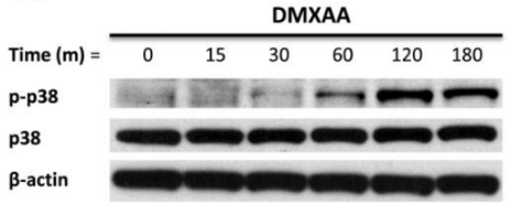
|
21819972 | |
| Growth inhibition assay | Cell proliferation |
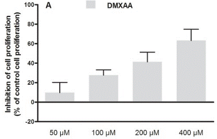
|
30138430 | |
| In Vivo | ||
| In Vivo | DMXAA treatment significantly protects C57BL/6J mice infected i.n. with 200 p.f.u. mouse-adapted H1N1 influenza PR8 virus with 60% survival, while the control group only exhibited 20% survival. [2] DMXAA significantly delays tumor growth induced by chemical carcinogen, increases the time to tumor doubling and increases time from treatment to euthanasia. After the treatment of DMXAA, median tumor doubling time, median tumour tripling time and median time from treatment to euthanasia in tumor-bearing animals are increased by approximately 4.4-, 1.8- and 2.7-fold, respectively. [4] | |
|---|---|---|
| 動物実験 | 動物モデル | Chemical carcinogen (NMU) is injected into female Wistar rats. |
| 投与量 | ≤300 mg/kg | |
| 投与経路 | Administered via i.p. | |
| NCT Number | Recruitment | Conditions | Sponsor/Collaborators | Start Date | Phases |
|---|---|---|---|---|---|
| NCT00856336 | Completed | Refractory Tumors |
Antisoma Research |
May 2003 | Phase 1 |
| NCT00863733 | Completed | Solid Tumors |
Cancer Research UK|Cancer Society Auckland |
May 1996 | Phase 1 |
化学情報
| 分子量 | 282.29 | 化学式 | C17H14O4 |
| CAS No. | 117570-53-3 | SDF | Download Vadimezan (DMXAA) SDFをダウンロードする |
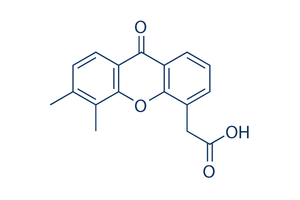
| Smiles | CC1=C(C2=C(C=C1)C(=O)C3=CC=CC(=C3O2)CC(=O)O)C | ||
| 保管 | |||
|
In vitro |
7.5%Sodium bicarbonate : 10 mg/mL DMSO : 7 mg/mL ( (24.79 mM); 吸湿したDMSOは溶解度を減少させます。新しいDMSOをご使用ください。) Water : Insoluble |
モル濃度計算器 |
|
in vivo Add solvents to the product individually and in order. |
投与溶液組成計算機 | ||||
実験計算
投与溶液組成計算機(クリア溶液)
ステップ1:実験データを入力してください。(実験操作によるロスを考慮し、動物数を1匹分多くして計算・調製することを推奨します)
mg/kg
g
μL
匹
ステップ2:投与溶媒の組成を入力してください。(ロット毎に適した溶解組成が異なる場合があります。詳細については弊社までお問い合わせください)
% DMSO
%
% Tween 80
% ddH2O
%DMSO
%
計算結果:
投与溶媒濃度: mg/ml;
DMSOストック溶液調製方法: mg 試薬を μL DMSOに溶解する(濃度 mg/mL, 注:濃度が当該ロットのDMSO溶解度を超える場合はご連絡ください。 )
投与溶媒調製方法:Take μL DMSOストック溶液に μL PEG300,を加え、完全溶解後μL Tween 80,を加えて完全溶解させた後 μL ddH2O,を加え完全に溶解させます。
投与溶媒調製方法:μL DMSOストック溶液に μL Corn oil,を加え、完全溶解。
注意:1.ストック溶液に沈殿、混濁などがないことをご確認ください;
2.順番通りに溶剤を加えてください。次のステップに進む前に溶液に沈殿、混濁などがないことを確認してから加えてください。ボルテックス、ソニケーション、水浴加熱など物理的な方法で溶解を早めることは可能です。
技術サポート
ストックの作り方、阻害剤の保管方法、細胞実験や動物実験の際に注意すべき点など、製品を取扱う時に問い合わせが多かった質問に対しては取扱説明書でお答えしています。
他に質問がある場合は、お気軽にお問い合わせください。
* 必須
Tags: Vadimezan (DMXAA)を買う | Vadimezan (DMXAA) ic50 | Vadimezan (DMXAA)供給者 | Vadimezan (DMXAA)を購入する | Vadimezan (DMXAA)費用 | Vadimezan (DMXAA)生産者 | オーダーVadimezan (DMXAA) | Vadimezan (DMXAA)化学構造 | Vadimezan (DMXAA)分子量 | Vadimezan (DMXAA)代理店
納期 国内在庫品:受注日の翌日(15時までの受注分) *北海道、九州、沖縄への配送は受注日より2日以上 を要する場合あり 海外在庫品:受注後1〜2週間