
- 阻害剤
- 研究分野別
- PI3K/Akt/mTOR
- Epigenetics
- Methylation
- Immunology & Inflammation
- Protein Tyrosine Kinase
- Angiogenesis
- Apoptosis
- Autophagy
- ER stress & UPR
- JAK/STAT
- MAPK
- Cytoskeletal Signaling
- Cell Cycle
- TGF-beta/Smad
- 化合物ライブラリー
- 抗体
- 新製品
- お問い合わせ
Pelitinib (EKB-569)
Pelitinib (EKB-569) is a potent irreversible EGFR inhibitor with IC50 of 38.5 nM. Pelitinib (EKB-569) also slightly inhibits Src, MEK/ERK and ErbB2 with IC50s of 282 nM, 800 nM and 1255 nM, respectively. Phase2.
CAS No. 257933-82-7
文献中Selleckの製品使用例(11)
カスタマーフィードバック3个实验数据
製品安全説明書
現在のバッチを見る:
純度:
99.49%
99.49
Pelitinib (EKB-569)関連製品
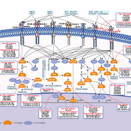
シグナル伝達経路
EGFR阻害剤の選択性比較
Cell Data
| Cell Lines | Assay Type | Concentration | Incubation Time | 活性情報 | PMID |
|---|---|---|---|---|---|
| Sf9 | Function assay | 10 mins | Inhibition of recombinant human His6-tagged EGFR cytoplasmic domain (645 to 1186 residues) expressed in baculovirus infected Sf9 insect cells assessed as reduction in autophosphorylation preincubated for 10 mins followed by ATP-MgCl2 addition and measured, IC50=0.0385μM. | 30600149 | |
| A431 | Function assay | 90 mins | Inhibition of EGFR in human A431 cells assessed as reduction in EGF-stimulated EGFR autophosphorylation preincuabted for 90 mins followed by EGF-stimulation by sandwich-ELISA, IC50=0.039μM. | 30973735 | |
| DiFi | Function assay | 2 hrs | Inhibition autophosphorylation of EGFR in human DiFi cells after 2 hrs by ELISA, IC50=0.07943μM. | 17689836 | |
| UCH1 | Antiproliferative assay | 72 hrs | Antiproliferative activity against human UCH1 cells measured after 72 hrs by alamar blue assay, IC50=0.09μM. | 30973735 | |
| UCH2 | Antiproliferative assay | 72 hrs | Antiproliferative activity against human UCH2 cells measured after 72 hrs by alamar blue assay, IC50=1.6μM. | 30973735 | |
| A431 | Function assay | Inhibition of EGFR autophosphorylation in human A431 cells, IC50=0.00802μM. | 20797871 | ||
| 他の多くの細胞株試験データをご覧になる場合はこちらをクリックして下さい | |||||
生物活性
| 製品説明 | Pelitinib (EKB-569) is a potent irreversible EGFR inhibitor with IC50 of 38.5 nM. Pelitinib (EKB-569) also slightly inhibits Src, MEK/ERK and ErbB2 with IC50s of 282 nM, 800 nM and 1255 nM, respectively. Phase2. | ||||||||||
|---|---|---|---|---|---|---|---|---|---|---|---|
| 特性 | An improved version of EKI-785. | ||||||||||
| Targets |
|
| In Vitro | ||||
| In vitro | Pelitinib displays much higher inhibitory activity against EGFR, compared with the closely related c-erbB-2, as well as other kinases such as Src, Cdk4, c-Met, Raf, and MEK/ERK, with IC50 ranging from 282 nM for Src to >20 μM for Cdk4. Consistently, Pelitinib treatment significantly inhibits the autophosphorylation of EGFR but not c-Met in A431 cells. [1] Pelitinib potently inhibits the proliferation of normal human keratinocytes (NHEK), as well as A431 and MDA-468 tumor cells with IC50 of 61 nM, 125 nM, and 260 nM, respectively, while displaying little activity against MCF-7 cells with IC50 of 3.6 μM. Pelitinib inhibits EGF-induced phosphorylation of EGFR in A431 and NHEK cells with IC50 of 20-80 nM, as well as the phosphorylation of STAT3 with IC50 of 30-70 nM. Pelitinib at 75-500 nM also specifically inhibits the activation of AKT and ERK1/2, without affecting NF-κB pathway. In NHEK cells, Pelitinib also potently inhibits TGF-α mediated EGFR activation with IC50 of 56 nM, as well as activation of STAT3 and ERK1/2 with IC50 of 60 nM and 62 nM, respectively. [2] | |||
|---|---|---|---|---|
| Kinase Assay | Autophosphorylation of EGFR in cells | |||
| For experiments using cells in culture, A431 cells are treated with various concentrations of Pelitinib for 2.75 hours before co-incubation with 100 ng/mL EGF for 0.25 hour. Cells are washed twice with cold phosphate-buffered saline (PBS) before adding to lysis buffer (10 mM Tris, pH 7.5, 5 mM ethylenediamine tetra-acetic acid (EDTA), 150 mM NaCl, 1% Triton X-100, 1% Sodium deoxycholate, 0.1 % SDS, 1 mM PMSF, 10 mg/mL pepstatin A, 10 mg/mL leupeptin, 20 KIU/mL aprotinin, 2 mM sodium orthovanadate, and 100 mM sodium fluoride) for 20 minutes on ice, before immunoprecipitation and SDS-PAGE-immunoblotting. For immunoprecipitation, cultured cells are placed in cold lysis buffer and immediately homogenized on ice with a polytron with several pulses. The homogenate is first centrifuged at 2500 rpm (20 minutes, 4 °C) and then again at 14,000 rpm in a microcentrifuge (10 minutes, 4 °C). Supernatants (1000 μg protein) are incubated for 2 hours at 4 °C with 15 mL of EGFR polyclonal antibody. After 2 hours, 50 μL of protein G plus/protein A agarose beads is added and incubated with constant rotation for 2 hours at 4 °C. After washing with lysis buffer, beads are boiled for 2 minutes in Laemmli sample buffer. Proteins are then resolved by SDS-PAGE, transferred to immobilon membrane and probed overnight with an anti-phosphotyrosine antibody conjugated with horseradish peroxidase (HRP). Membranes are developed using the ECL reagent. Total EGFR protein is determined by stripping membranes and re-probing with receptor-specific antibodies. Quantitation of bands is done by densitometry, using ImageQuant software with a Molecular Dynamics laser transmittance scanner. | ||||
| 細胞実験 | 細胞株 | NHEK, A431, MCF-7, and MDA-468 | ||
| 濃度 | Dissolved in DMSO, final concentrations ~10 μM | |||
| 反応時間 | 5 days | |||
| 実験の流れ | Cells are seeded in 96-well dishes, and after 2 hours, Pelitinib is added and incubated for 5 days. After incubation, the medium is removed from each well and fresh medium (150 μL) + 1 mg/mL MTT solution (50 μL) is added. After incubation for 2 hours at 37 °C, the medium is replaced with 150 μL DMSO, and absorbance at 540 nm in each well is determined. The IC50 is calculated by linear regression of the data. |
|||
| In Vivo | ||
| In Vivo | A single oral dose of 10 mg/kg Pelitinib potently inhibits the EGFR phosphorylation in A431 xenografts with over-expressed EGFR, by 90% within 1 hour, and by >50% after 24 hours. Administration of Pelitinib at 20 mg/kg/day inhibits tumorigenesis in APCMin/+ mice by 87%, equivalent to the effect of used with 2 times doses of EKI-785 (40 mg/kg/day), consistent with greater in vivo potency. [1] Pelitinib selectively inhibits EGFR signaling in airway epithelial cells in vivo. In the mouse model of airway epithelial remodeling that is inducible by viral infection and features a delayed but permanent switch to goblet cell metaplasia, Pelitinib treatment at 20 mg/kg/day corrects all 3 aspects of epithelial remodeling, by completely blocking the increase of ciliated cells and decrease of Clara cells, and significantly inhibiting the metaplasia of goblet cells. [3] | |
|---|---|---|
| 動物実験 | 動物モデル | Athymic nu/nu female mice bearing subcutaneous A431 tumors, or APCMin/+ male mice, a murine model of human familial adenomatous polyposis (FAP) |
| 投与量 | 10, or 20 mg/kg/day | |
| 投与経路 | Oral gavage | |
化学情報
| 分子量 | 467.92 | 化学式 | C24H23ClFN5O2 |
| CAS No. | 257933-82-7 | SDF | Download Pelitinib (EKB-569) SDFをダウンロードする |
| Smiles | CCOC1=C(C=C2C(=C1)N=CC(=C2NC3=CC(=C(C=C3)F)Cl)C#N)NC(=O)C=CCN(C)C | ||
| 保管 | |||
|
In vitro |
DMSO : 13 mg/mL ( (27.78 mM); 吸湿したDMSOは溶解度を減少させます。新しいDMSOをご使用ください。) Water : Insoluble Ethanol : Insoluble |
モル濃度計算器 |
|
in vivo Add solvents to the product individually and in order. |
投与溶液組成計算機 | ||||
実験計算
投与溶液組成計算機(クリア溶液)
ステップ1:実験データを入力してください。(実験操作によるロスを考慮し、動物数を1匹分多くして計算・調製することを推奨します)
mg/kg
g
μL
匹
ステップ2:投与溶媒の組成を入力してください。(ロット毎に適した溶解組成が異なる場合があります。詳細については弊社までお問い合わせください)
% DMSO
%
% Tween 80
% ddH2O
%DMSO
%
計算結果:
投与溶媒濃度: mg/ml;
DMSOストック溶液調製方法: mg 試薬を μL DMSOに溶解する(濃度 mg/mL, 注:濃度が当該ロットのDMSO溶解度を超える場合はご連絡ください。 )
投与溶媒調製方法:Take μL DMSOストック溶液に μL PEG300,を加え、完全溶解後μL Tween 80,を加えて完全溶解させた後 μL ddH2O,を加え完全に溶解させます。
投与溶媒調製方法:μL DMSOストック溶液に μL Corn oil,を加え、完全溶解。
注意:1.ストック溶液に沈殿、混濁などがないことをご確認ください;
2.順番通りに溶剤を加えてください。次のステップに進む前に溶液に沈殿、混濁などがないことを確認してから加えてください。ボルテックス、ソニケーション、水浴加熱など物理的な方法で溶解を早めることは可能です。
技術サポート
ストックの作り方、阻害剤の保管方法、細胞実験や動物実験の際に注意すべき点など、製品を取扱う時に問い合わせが多かった質問に対しては取扱説明書でお答えしています。
他に質問がある場合は、お気軽にお問い合わせください。
* 必須
Tags: Pelitinib (EKB-569)を買う | Pelitinib (EKB-569) ic50 | Pelitinib (EKB-569)供給者 | Pelitinib (EKB-569)を購入する | Pelitinib (EKB-569)費用 | Pelitinib (EKB-569)生産者 | オーダーPelitinib (EKB-569) | Pelitinib (EKB-569)化学構造 | Pelitinib (EKB-569)分子量 | Pelitinib (EKB-569)代理店
納期 国内在庫品:受注日の翌日(15時までの受注分) *北海道、九州、沖縄への配送は受注日より2日以上 を要する場合あり 海外在庫品:受注後1〜2週間