
- 阻害剤
- 研究分野別
- PI3K/Akt/mTOR
- Epigenetics
- Methylation
- Immunology & Inflammation
- Protein Tyrosine Kinase
- Angiogenesis
- Apoptosis
- Autophagy
- ER stress & UPR
- JAK/STAT
- MAPK
- Cytoskeletal Signaling
- Cell Cycle
- TGF-beta/Smad
- 化合物ライブラリー
- 抗体
- 新製品
- お問い合わせ
ICG-001
ICG-001 antagonizes Wnt/β-catenin/TCF-mediated transcription and specifically binds to CREB-binding protein (CBP) with IC50 of 3 μM, but is not the related transcriptional coactivator p300. ICG-001 induces apoptosis.
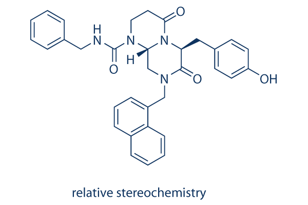
CAS No. 780757-88-2 (relative stereochemistry); 847591-62-2 (absolute stereochemistry)
文献中Selleckの製品使用例(182)
製品安全説明書
現在のバッチを見る:
純度:
99.75%
99.75
ICG-001と併用されることが多い化合物
Foscenvivint and Auranofin synergistically inhibit the growth of colon cancer in subcutaneous xenograft mice models and restrain metastasis in lung metastasis mice models.
Foscenvivint ameliorates Paclitaxel-induced increase in FOXM1 expression and cancer stem cells (CSC) phenotype as well as tumor initiation capacity in vitro.
Foscenvivint and JQ1 combination treatment induces strong cytotoxic effects in H3.3K27M-mutated diffuse intrinsic pontine gliomas (DIPG) cell lines.
Foscenvivint and Pyrvinium plus Bortezomib decrease cell viability and increase cell apoptosis rate in RPMI-8226BR and KMS-11BR cell lines.
Foscenvivint and Wnt-C59 inhibit in vitro growth of human cholangiocarcinoma cells and reduce tumor area and number in xenograft and thioacetamide models.
ICG-001関連製品
シグナル伝達経路
Epigenetic Reader Domain阻害剤の選択性比較
Cell Data
| Cell Lines | Assay Type | Concentration | Incubation Time | 活性情報 | PMID |
|---|---|---|---|---|---|
| HK-2 | Function Assay | 10 µM | 3 h | reduced the expression of TGF-β1, α-SMA, and CTGF after treatment with HHE | 23690997 |
| HKC-8 | Function Assay | 10 µM | 24 h | abolishes β-catenin–mediated RAS induction | 25012166 |
| SH-SY5Y | Apoptosis Assay | 10 μM | 24 h | inhibits the neuroprotective effects of hypoxia against PrP (106-126)-mediated neuronal cell death | 23900566 |
| L3.6pl | Growth Inhibition Assay | 1-20 μM | 2/4/6 d | inhibits the cell growth in a dose-dependent manner | 25082960 |
| PANC-1 | Growth Inhibition Assay | 1-20 μM | 2/4/6 d | inhibits the cell growth in a dose-dependent manner | 25082960 |
| MiaPaCa-2 | Growth Inhibition Assay | 1-20 μM | 2/4/6 d | inhibits the cell growth in a dose-dependent manner | 25082960 |
| AsPC-1 | Growth Inhibition Assay | 1-20 μM | 2/4/6 d | inhibits the cell growth in a dose-dependent manner | 25082960 |
| SH-SY5Y | Apoptosis Assay | 50 μm | 24 h | blocks the protective effect of melatonin against PrP (106–126)-induced apoptotic signals | 25251028 |
| HepT1 | Apoptosis Assay | 0-100 μM | 24 h | IC50=34 μM | 23266718 |
| HuH6 | Apoptosis Assay | 0-100 μM | 24 h | IC50=39 μM | 23266718 |
| RLE-6TN | Function Assay | 2.5/5/7.5 μM | 48 h | inhibits TGF-β1-induced α-SMA induction and EMT | 22241478 |
| HKC-8 | Function Assay | 5/10/20 μM | 48 h | blocks β-catenin-driven gene expression | 21816937 |
| LoVo | Cytotoxicity assay | 10 uM | 72 hrs | Cytotoxicity against Wnt/beta-catenin signalling dependent human LoVo cells assessed as cell viability at 10 uM after 72 hrs by ATPlite assay | ChEMBL |
| NCI-H1703 | Function assay | 10 uM | 24 hrs | Inhibition of TNIK in human NCI-H1703 cells transfected with lentiviral vector 7TFP assessed as reduction of GSK3 inhibitor X activated TNIK-mediated Wnt/TCF/beta-catenin-dependent transcription at 10 uM after 24 hrs by luciferase reporter assay | ChEMBL |
| HCT116 | Cytotoxicity assay | 10 uM | 72 hrs | Cytotoxicity against Wnt/beta-catenin signalling dependent human HCT116 cells assessed as cell viability at 10 uM after 72 hrs by ATPlite assay | ChEMBL |
| MCF7 | Function Assay | 5 μm | inhibits leptin-mediated increased expression of Snail, Slug, and Zeb2 | 22270359 | |
| SW480 | Growth Inhibition Assay | 2-100 μM | IC50=5.8±0.68 μM | 15782138 | |
| A549 | Antiproliferative assay | 72 hrs | Antiproliferative activity against human A549 cells after 72 hrs by MTT assay, GI50 = 6.1 μM. | 24950489 | |
| HepG2 | Antiproliferative assay | 72 hrs | Antiproliferative activity against human HepG2 cells after 72 hrs by MTT assay, GI50 = 12.7 μM. | 24950489 | |
| LoVo | Antiproliferative assay | 72 hrs | Antiproliferative activity against human LoVo cells after 72 hrs by MTT assay, GI50 = 15.6 μM. | 24950489 | |
| HT-29 | Antiproliferative assay | 72 hrs | Antiproliferative activity against human HT-29 cells after 72 hrs by MTT assay, GI50 = 17.2 μM. | 24950489 | |
| HT29 | Function assay | 24 hrs | Inhibition of Wnt signaling in human HT29 cells assessed as inhibition of beta-catenin-mediated Tcf/Lef transcriptional activity after 24 hrs by dual luciferase reporter gene assay relative to control, IC50 = 18.7 μM. | 24950489 | |
| SW480 | Function assay | Inhibition of CBP binding to beta-casein in human SW480 cells by immunoblot analysis, IC50 = 1.3 μM. | 23232060 | ||
| TC32 | qHTS assay | qHTS of pediatric cancer cell lines to identify multiple opportunities for drug repurposing: Primary screen for TC32 cells | 29435139 | ||
| A673 | qHTS assay | qHTS of pediatric cancer cell lines to identify multiple opportunities for drug repurposing: Primary screen for A673 cells | 29435139 | ||
| DAOY | qHTS assay | qHTS of pediatric cancer cell lines to identify multiple opportunities for drug repurposing: Primary screen for DAOY cells | 29435139 | ||
| BT-37 | qHTS assay | qHTS of pediatric cancer cell lines to identify multiple opportunities for drug repurposing: Primary screen for BT-37 cells | 29435139 | ||
| RD | qHTS assay | qHTS of pediatric cancer cell lines to identify multiple opportunities for drug repurposing: Primary screen for RD cells | 29435139 | ||
| MG 63 (6-TG R) | qHTS assay | qHTS of pediatric cancer cell lines to identify multiple opportunities for drug repurposing: Primary screen for MG 63 (6-TG R) cells | 29435139 | ||
| NB1643 | qHTS assay | qHTS of pediatric cancer cell lines to identify multiple opportunities for drug repurposing: Primary screen for NB1643 cells | 29435139 | ||
| OHS-50 | qHTS assay | qHTS of pediatric cancer cell lines to identify multiple opportunities for drug repurposing: Primary screen for OHS-50 cells | 29435139 | ||
| SJ-GBM2 | qHTS assay | qHTS of pediatric cancer cell lines to identify multiple opportunities for drug repurposing: Primary screen for SJ-GBM2 cells | 29435139 | ||
| SK-N-MC | qHTS assay | qHTS of pediatric cancer cell lines to identify multiple opportunities for drug repurposing: Primary screen for SK-N-MC cells | 29435139 | ||
| NB-EBc1 | qHTS assay | qHTS of pediatric cancer cell lines to identify multiple opportunities for drug repurposing: Primary screen for NB-EBc1 cells | 29435139 | ||
| LAN-5 | qHTS assay | qHTS of pediatric cancer cell lines to identify multiple opportunities for drug repurposing: Primary screen for LAN-5 cells | 29435139 | ||
| 他の多くの細胞株試験データをご覧になる場合はこちらをクリックして下さい | |||||
生物活性
| 製品説明 | ICG-001 antagonizes Wnt/β-catenin/TCF-mediated transcription and specifically binds to CREB-binding protein (CBP) with IC50 of 3 μM, but is not the related transcriptional coactivator p300. ICG-001 induces apoptosis. | ||
|---|---|---|---|
| Targets |
|
| In Vitro | ||||
| In vitro |
ICG-001 has no effect on the related reporter construct, FOPFLASH, which contains mutated TCF sites. After treatment with 25μM of ICG-001 for 8 hours, SW480 cell reduces the steady-state levels of Survivin and Cyclin D1 RNA and protein, both of which can be up-regulated by β-catenin. ICG-001 selectively induces apoptosis in transformed cells but not in normal colon cells, reduces in vitro growth of colon carcinoma cells. [1] ICG-001, can phenotypically rescue normal nerve growth factor (NGF) -induced neuronal differentiation and neurite outgrowth in the presenilin-1 mutant cells, emphasizing the importance of the TCF/β-catenin signaling pathway on neurite outgrowth and neuronal differentiation. [2] A recent study demonstrates that 5μM ICG-001 inhibits leptin-induced EMT, invasion and tumorsphere formation in MCF7 cells. [3] |
|||
|---|---|---|---|---|
| Kinase Assay | DUAL-Luciferase Reporter Assay | |||
| The Dual-Luciferase Reporter (DLR) Assay System provides an efficient means of performing dual reporter assays. In the DLRTM Assay, the activities of firefly (Photinus pyralis) and Renilla (Renilla reniformis, also known as sea pansy) luciferases are measured sequentially from a single sample. The firefly luciferase reporter is measured first by adding Luciferase Assay Reagent II (LAR II) to generate a “glow-type” luminescent signal. After quantifying the firefly luminescence, this reaction is quenched, and the Renilla luciferase reaction is initiated by simultaneously adding Stop & Glo® Reagent to the same tube. The Stop & Glo® Reagent also produces a “glow-type” signal from the Renilla luciferase, which decays slowly over the course of the measurement. In the DLRTM Assay System, both reporters yield linear assays with subattomole (<10-18) sensitivities and no endogenous activity of either reporter in the experimental host cells. Furthermore, the integrated format of the DLRTM Assay provides rapid quantitation of both reporters either in transfected cells or in cell-free transcription/translation reactions. | ||||
| 細胞実験 | 細胞株 | Human colon carcinoma cell lines SW480, SW620, and HCT116, normal colonic epithelial cell line CCD-841Co | ||
| 濃度 | ~25 μM | |||
| 反応時間 | 24 hours | |||
| 実験の流れ | 1. Prior to starting the assay, prepare the Apo-ONE Caspase-3/7 Reagent, and mix thoroughly. 2. For best results, empirical determination of the optimal cell number, apoptosis induction treatment and incubation period for the cell culture system may be necessary. 3. Use identical cell numbers and volumes for the assay and the negative control samples. 4. Do not mix Apo-ONE Caspase-3/7 Reagent and samples by manual pipetting. Mixing in this manner is unnecessary and may create bubbles that interfere with fluorescence readings or cross-contaminate the samples. Gentle mixing may be performed using a plate shaker. 5. Total incubation time for the assay depends upon the amount of caspase- 3/7 present in the sample. 6. The Apo-ONE Caspase-3/7 Reagent is formulated to mediate cellular lysis and support optimal caspase-3/7 activity. In rare instances, the reagent does not affect complete lysis of cultured cells. In such cases, lysis is enhanced by a freeze-thaw cycle. For best results, freeze at -70 °C, then thaw at room temperature. After equilibration, mix to homogeneity and incubate until measurable fluorescence is achieved |
|||
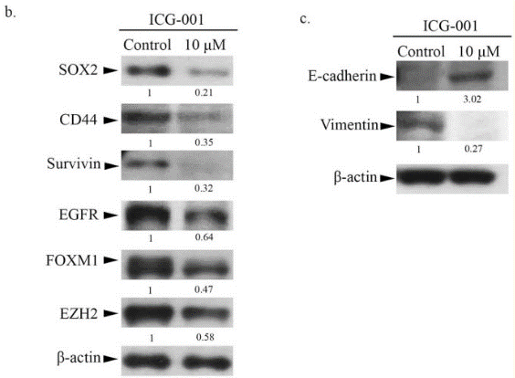
| 実験結果図 | Methods | Biomarkers | 結果図 | PMID |
| Western blot | SOX-2 / CD44 / Survivin / EGFR / FOXM1 / EZH2 / Vimentin pβ-catenin beta-catenin / F-actin ABC / Beta-catenin / E-cadherin / N-cadherin / MMP-9 / c-MYC / Actin / Histone H3 CCNB1 / Cyclin D1 |
|
25897700 | |
| In Vivo | ||
| In Vivo |
Administration of a water-soluble analog of ICG-001 for 9 weeks reduces the formation of colon and small intestinal polyps by 42% as effectively as the nonsteroidal antiinflammatory agent Sulindac, which has consistently demonstrated efficacy in this model. No overt toxicity is detected throughout the course of treatment. In the SW620 nude mouse xenograft model of tumor regression, 150 mg/kg, i.v. of analog demonstrates a dramatic reduction in tumor volume over the 19-day course of treatment, with no mortality or weight loss. [1] ICG-001 (5 mg/kg per day) significantly inhibits beta-catenin signaling and attenuates bleomycin-induced lung fibrosis in mice, while concurrently preserving the epithelium. [4] |
|
|---|---|---|
| 動物実験 | 動物モデル | C57BL/6 and nude mice tumor models |
| 投与量 | 20 mg/ kg | |
| 投与経路 | i.p. | |
化学情報
| 分子量 | 548.63 | 化学式 | C33H32N4O4 |
| CAS No. | 780757-88-2 (relative stereochemistry); 847591-62-2 (absolute stereochemistry) | SDF | Download ICG-001 SDFをダウンロードする |
| Smiles | C1CN(C2CN(C(=O)C(N2C1=O)CC3=CC=C(C=C3)O)CC4=CC=CC5=CC=CC=C54)C(=O)NCC6=CC=CC=C6 | ||
| 保管 | |||
|
In vitro |
DMSO : 30 mg/mL ( (54.68 mM); 吸湿したDMSOは溶解度を減少させます。新しいDMSOをご使用ください。) Water : Insoluble Ethanol : Insoluble |
モル濃度計算器 |
|
in vivo Add solvents to the product individually and in order. |
投与溶液組成計算機 | ||||
実験計算
投与溶液組成計算機(クリア溶液)
ステップ1:実験データを入力してください。(実験操作によるロスを考慮し、動物数を1匹分多くして計算・調製することを推奨します)
mg/kg
g
μL
匹
ステップ2:投与溶媒の組成を入力してください。(ロット毎に適した溶解組成が異なる場合があります。詳細については弊社までお問い合わせください)
% DMSO
%
% Tween 80
% ddH2O
%DMSO
%
計算結果:
投与溶媒濃度: mg/ml;
DMSOストック溶液調製方法: mg 試薬を μL DMSOに溶解する(濃度 mg/mL, 注:濃度が当該ロットのDMSO溶解度を超える場合はご連絡ください。 )
投与溶媒調製方法:Take μL DMSOストック溶液に μL PEG300,を加え、完全溶解後μL Tween 80,を加えて完全溶解させた後 μL ddH2O,を加え完全に溶解させます。
投与溶媒調製方法:μL DMSOストック溶液に μL Corn oil,を加え、完全溶解。
注意:1.ストック溶液に沈殿、混濁などがないことをご確認ください;
2.順番通りに溶剤を加えてください。次のステップに進む前に溶液に沈殿、混濁などがないことを確認してから加えてください。ボルテックス、ソニケーション、水浴加熱など物理的な方法で溶解を早めることは可能です。
技術サポート
ストックの作り方、阻害剤の保管方法、細胞実験や動物実験の際に注意すべき点など、製品を取扱う時に問い合わせが多かった質問に対しては取扱説明書でお答えしています。
他に質問がある場合は、お気軽にお問い合わせください。
* 必須
よくある質問(FAQ)
質問1:
If the compound is stored in DMSO at -80, how long would it be stable? For cell culture, how long should I change for the fresh medium with ICG-001?
回答
The product in DMSO solution can be stored at 4 degree for 1 week and -20 degree for 1 month. The best storage condition is solid powder, even at -80 the solution is not stable enough for long term storage. For cell culture, you need change medium every 48h.
Tags: ICG-001を買う | ICG-001 ic50 | ICG-001供給者 | ICG-001を購入する | ICG-001費用 | ICG-001生産者 | オーダーICG-001 | ICG-001化学構造 | ICG-001分子量 | ICG-001代理店
納期 国内在庫品:受注日の翌日(15時までの受注分) *北海道、九州、沖縄への配送は受注日より2日以上 を要する場合あり 海外在庫品:受注後1〜2週間