
- 阻害剤
- 研究分野別
- PI3K/Akt/mTOR
- Epigenetics
- Methylation
- Immunology & Inflammation
- Protein Tyrosine Kinase
- Angiogenesis
- Apoptosis
- Autophagy
- ER stress & UPR
- JAK/STAT
- MAPK
- Cytoskeletal Signaling
- Cell Cycle
- TGF-beta/Smad
- 化合物ライブラリー
- 抗体
- 新製品
- お問い合わせ
PAC-1
PAC-1 is a potent procaspase-3 activator with EC50 of 0.22 μM and the first small molecule known to directly activate procaspase-3 to caspase-3.
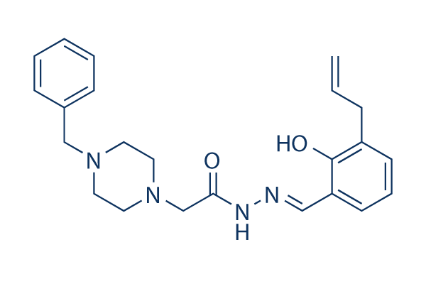
CAS No. 315183-21-2
文献中Selleckの製品使用例(16)
カスタマーフィードバック1个实验数据
製品安全説明書
現在のバッチを見る:
純度:
99.99%
99.99
PAC-1関連製品
シグナル伝達経路
Caspase阻害剤の選択性比較
生物活性
| 製品説明 | PAC-1 is a potent procaspase-3 activator with EC50 of 0.22 μM and the first small molecule known to directly activate procaspase-3 to caspase-3. | ||
|---|---|---|---|
| 特性 | The first small molecule known to directly activate procaspase-3 to caspase-3. | ||
| Targets |
|
| In Vitro | ||||
| In vitro | PAC-1 activates procaspase-7 in a less efficient manner with EC50 of 4.5 μM. Elevated caspase 3 level in cancer cell lines allows PAC-1 to selectively induce apoptosis in a manner proportional to procaspase-3 concentration with IC50 of 0.35 μM for NCI-H226 cells to ~3.5 μM for UACC-62 cells. PAC-1 induces apoptosis in the primary cancerous cells with IC50 values of 3 nM to 1.41 μM, more potently than in the adjacent noncancerous cells with IC50 of 5.02 μM to 9.98 μM, which is also directly related to the distinct procaspase-3 concentration. [1] PAC-1 activates procaspase-3 by chelating zinc ions, thus relieving the zinc-mediated inhibition and allowing procaspase-3 to auto-activate itself to caspase-3. [2] PAC-1 is capable to induce cell death in Bax/Bak double-knockout cells and Bcl-2 and Bcl-xL-overexpressing cells with the same efficacy as its wild-type counterpart in a delayed manner. PAC-1 induces cytochrome c release in a caspase-3 independent manner, which subsequently triggers downstream caspase-3 activation and cell death. PAC-1 can not induce cell death and caspase-3 activation in Apaf-1 knockout cells, suggesting that apoptosome formation is essential for caspase-3 activation by PAC-1-mediated cell death. [3] | |||
|---|---|---|---|---|
| Kinase Assay | In vitro procaspase-3 activation | |||
| Procaspase-3 is expressed and purified in Escherichia coli. Various concentrations of PAC-1 are added to 90 μL of a 50 ng/mL of procaspase-3 in caspase assay buffer in a 96-well plate, The plate is incubated for 12 hours at 37 °C. A 10 μL volume of a 2 mM solution of caspase-3 peptidic substrate acetyl Asp-Glu-Val-Asp-p-nitroanilide (Ac-DEVD-pNa) in caspase assay buffer is then added to each well. The plate is read every 2 minutes at 405 nm for 2 hours in a Spectra Max Plus 384 well plate reader. The slope of the linear portion for each well is determined, and the relative increase in activation from untreated control wells is calculated. | ||||
| 細胞実験 | 細胞株 | U-937, HL-60, CRL-1872, ACHN, NCI-H226, Hs888Lu, Hs578Bst, MCF-10A, SK-MEL-5, BT-20, MDA-MB-231, UACC-62, SK-N-SH, B16-F10 , Hs 578t, and PC-12 | ||
| 濃度 | Dissolved in DMSO, final concentrations ~100 μM | |||
| 反応時間 | 72 hours | |||
| 実験の流れ | Cells are exposed to various concentrations of PAC-1 for 72 hours. Cell death is quantified by the addition of MTS/PMS CellTiter 96 Cell Proliferation Assay reagent. The plates are incubated at 37 °C for approximately 1 hour (until the colored product formed), and the absorbance is measured at 490 n | |||
| 実験結果図 | Methods | Biomarkers | 結果図 | PMID |
| Western blot | Ero1-Lα / Calnexin / IRE-1α / p-eIF2α / eIF2α / CHOP |
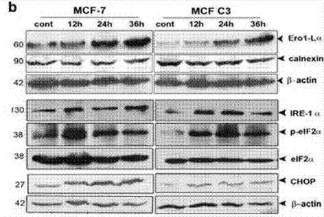
|
24357799 | |
| Immunofluorescence | Hif1α p-H2AX / Rad51 |
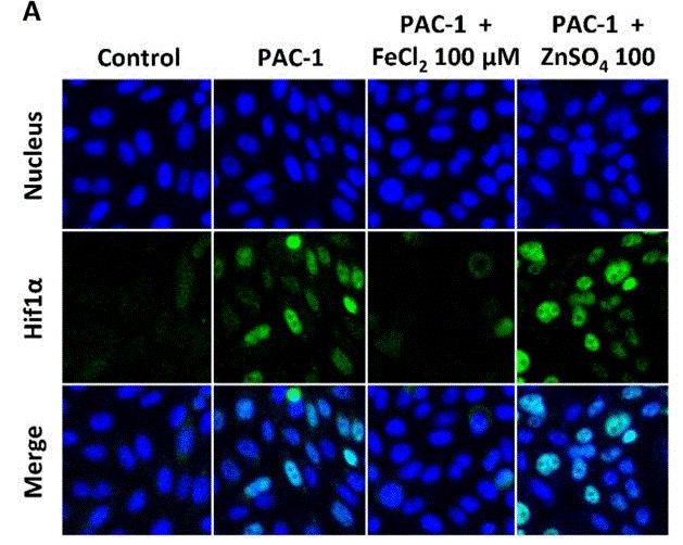
|
30287840 | |
| In Vivo | ||
| In Vivo | Administration of PAC-1 at 5 mg with low and steady releasing significantly inhibits the growth of ACHN renal cancer xenograft in mice. Oral administration of PAC-1 (50 or 100 mg/kg) significantly retards tumor growth of NCI-H226 lung cancer xenograft in a dose-dependent manner, and markedly prevents the cancer cells from infiltrating the lung tissue. The in vivo anti-tumor effect of PAC-1 is ascribed to procaspase-3 activation and subsequently apoptosis induction consistent with the activity in vitro. [1] | |
|---|---|---|
| 動物実験 | 動物モデル | Ovariectomized female athymic BALB/c (nude, nu/nu) mice injected subcutaneously with ACHN cells, male athymic BALB/c nude mice injected subcutaneously with NCI-H226 cells, and male athymic BALB/c–/– mice injected intravenously with NCI-H226 cells |
| 投与量 | ~100 mg/kg | |
| 投与経路 | Pellet implantation subcutaneously or oral gavage | |
| NCT Number | Recruitment | Conditions | Sponsor/Collaborators | Start Date | Phases |
|---|---|---|---|---|---|
| NCT05967741 | Recruiting | Platelet Aggregation Spontaneous|Vascular Thrombosis |
University of California Davis |
July 20 2023 | Not Applicable |
| NCT03927248 | Withdrawn | Metastatic Renal Cell Carcinoma |
HealthPartners Institute |
September 2020 | Phase 1|Phase 2 |
| NCT03441412 | Completed | Healthy Volunteers |
Vastra Gotaland Region|Gothia Forum - Center for Clinical Trial|Uppsala University |
February 28 2018 | Phase 1 |
化学情報
| 分子量 | 392.49 | 化学式 | C23H28N4O2 |
| CAS No. | 315183-21-2 | SDF | Download PAC-1 SDFをダウンロードする |
| Smiles | C=CCC1=C(C(=CC=C1)C=NNC(=O)CN2CCN(CC2)CC3=CC=CC=C3)O | ||
| 保管 | |||
|
In vitro |
DMSO : 78 mg/mL ( (198.73 mM); 吸湿したDMSOは溶解度を減少させます。新しいDMSOをご使用ください。) Ethanol : 78 mg/mL Water : Insoluble |
モル濃度計算器 |
|
in vivo Add solvents to the product individually and in order. |
投与溶液組成計算機 | ||||
実験計算
投与溶液組成計算機(クリア溶液)
ステップ1:実験データを入力してください。(実験操作によるロスを考慮し、動物数を1匹分多くして計算・調製することを推奨します)
mg/kg
g
μL
匹
ステップ2:投与溶媒の組成を入力してください。(ロット毎に適した溶解組成が異なる場合があります。詳細については弊社までお問い合わせください)
% DMSO
%
% Tween 80
% ddH2O
%DMSO
%
計算結果:
投与溶媒濃度: mg/ml;
DMSOストック溶液調製方法: mg 試薬を μL DMSOに溶解する(濃度 mg/mL, 注:濃度が当該ロットのDMSO溶解度を超える場合はご連絡ください。 )
投与溶媒調製方法:Take μL DMSOストック溶液に μL PEG300,を加え、完全溶解後μL Tween 80,を加えて完全溶解させた後 μL ddH2O,を加え完全に溶解させます。
投与溶媒調製方法:μL DMSOストック溶液に μL Corn oil,を加え、完全溶解。
注意:1.ストック溶液に沈殿、混濁などがないことをご確認ください;
2.順番通りに溶剤を加えてください。次のステップに進む前に溶液に沈殿、混濁などがないことを確認してから加えてください。ボルテックス、ソニケーション、水浴加熱など物理的な方法で溶解を早めることは可能です。
技術サポート
ストックの作り方、阻害剤の保管方法、細胞実験や動物実験の際に注意すべき点など、製品を取扱う時に問い合わせが多かった質問に対しては取扱説明書でお答えしています。
他に質問がある場合は、お気軽にお問い合わせください。
* 必須
Tags: PAC-1を買う | PAC-1 ic50 | PAC-1供給者 | PAC-1を購入する | PAC-1費用 | PAC-1生産者 | オーダーPAC-1 | PAC-1化学構造 | PAC-1分子量 | PAC-1代理店
納期 国内在庫品:受注日の翌日(15時までの受注分) *北海道、九州、沖縄への配送は受注日より2日以上 を要する場合あり 海外在庫品:受注後1〜2週間