
- 阻害剤
- 研究分野別
- PI3K/Akt/mTOR
- Epigenetics
- Methylation
- Immunology & Inflammation
- Protein Tyrosine Kinase
- Angiogenesis
- Apoptosis
- Autophagy
- ER stress & UPR
- JAK/STAT
- MAPK
- Cytoskeletal Signaling
- Cell Cycle
- TGF-beta/Smad
- 化合物ライブラリー
- 抗体
- 新製品
- お問い合わせ
Stattic
Stattic, the first nonpeptidic small molecule, potently inhibits STAT3 activation and nuclear translocation with IC50 of 5.1 μM in cell-free assays, highly selectivity over STAT1. Stattic induces apoptosis.
CAS No. 19983-44-9
文献中Selleckの製品使用例(238)
製品安全説明書
現在のバッチを見る:
純度:
99.88%
99.88
Stattic関連製品
| 関連ターゲット | STAT1 STAT3 STAT5 STAT6 | もっと見る |
|---|---|---|
| 関連化合物ライブラリー | Kinase Inhibitor Library FDA-approved Drug Library Natural Product Library Tyrosine Kinase Inhibitor Library JAK/STAT compound library | もっと見る |
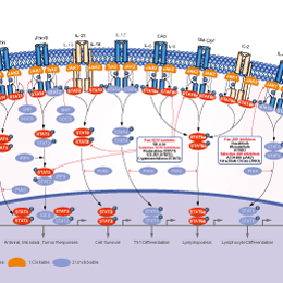
シグナル伝達経路
STAT阻害剤の選択性比較
Cell Data
| Cell Lines | Assay Type | Concentration | Incubation Time | 活性情報 | PMID |
|---|---|---|---|---|---|
| CNE2 | Function Assay | 0-20 μM | 0-4 h | inhibits Stat3 activation in a dose- and time-dependent manner | 23382914 |
| CNE1 | Function Assay | 0-20 μM | 0-4 h | inhibits Stat3 activation in a dose- and time-dependent manner | 23382914 |
| HONE1 | Function Assay | 20 µM | 48 h | blocks the IL-6 increased phosphorylation of Stat3 | 23382914 |
| CNE2 | Function Assay | 20 µM | 48 h | blocks the IL-6 increased phosphorylation of Stat3 | 23382914 |
| CNE1 | Function Assay | 20 µM | 48 h | blocks the IL-6 increased phosphorylation of Stat3 | 23382914 |
| T24 | Function Assay | 2/10/20 μM | 24 h | causes dose-dependent inhibition of the CXCL12-induced increase of invading cells | 23526079 |
| SW837 | Function Assay | 2.5/10 μM | 30 min | sensitizes cells to chemoradiotherapy in a dose-dependent manner | 23934972 |
| W480 | Function Assay | 2.5/10 μM | 30 min | sensitizes cells to chemoradiotherapy in a dose-dependent manner | 23934972 |
| OV2008 | Apoptosis Assay | 0-10 μM | 24/48 h | induces apoptosis in a dose and time dependent manner | 23962558 |
| C13* | Apoptosis Assay | 0-10 μM | 24/48 h | induces apoptosis in a dose and time dependent manner | 23962558 |
| HTR8/SVneo | Function Assay | 1 μM | 48 h | significantly increases migration by OSM | 24060241 |
| HTR8/SVneo | Function Assay | 0.5/1 μM | 48 h | restores the expression of E-cadherin suppressed by OSM | 24060241 |
| HTR8/SVneo | Function Assay | 1 μM | 1 h | suppressed OSM-induced STAT3 phosphorylation | 24060241 |
| HMECs | Function Assay | 10 μM | 2 h | inhibits IFNα mediated phosphorylation of STAT1, STAT2 and STAT3 | 24211327 |
| MCF7-HER2 | Growth Inhibition Assay | 5 μM | 24 h | enhances cell growth inhibition combined with Herceptin | 24297508 |
| MCF7-HER2 | Function Assay | 5 μM | 24 h | decreases the expression levels of EMT markers, vimentin and slug | 24297508 |
| MCF7-HER2 | Function Assay | 5 μM | 24 h | diminishes Sox-2, Oct-4, and slug expression | 24297508 |
| MCF7-HER2 | Growth Inhibition Assay | 0-10 μM | 48 h | induces cell death dose dependently | 24297508 |
| SK-BR-3 | Function Assay | 10 µM | 24 h | reduces P-STAT3 expression | 24376586 |
| SUM-159 | Function Assay | 10 µM | 24 h | reduces P-STAT3 expression | 24376586 |
| MDA-MB-231 | Function Assay | 10 µM | 24 h | reduces P-STAT3 expression | 24376586 |
| HaCaT | Apoptosis Assay | 10 µM | 20 min | enhances the apoptotic effects of everolimus | 24423131 |
| HaCaT | Growth Inhibition Assay | 10 µM | 20 min | enhances everolimus-induced cell growth inhibition | 24423131 |
| MCF-7/LCC9 | Growth Inhibition Assay | 0.469-3.75 μM | 5 d | reduces cell number significantly | 24728078 |
| MCF-7/LCC1 | Growth Inhibition Assay | 0.469-3.75 μM | 5 d | reduces cell number significantly | 24728078 |
| MCF-7 | Growth Inhibition Assay | 0.469-3.75 μM | 5 d | reduces cell number significantly | 24728078 |
| CD4+ | Apoptosis Assay | 10 μm | 24 h | induces apoptosis strongly | 24756111 |
| HuT-78 | Cell Viability Assay | 1-10 μM | 72 h | causes a dose-dependent inhibition of the viability | 24756111 |
| SeAx | Cell Viability Assay | 1-10 μM | 72 h | causes a dose-dependent inhibition of the viability | 24756111 |
| SS | Cell Viability Assay | 1-10 μM | 72 h | causes a dose-dependent inhibition of the viability | 24756111 |
| ELL-primed hNSCs | Cell Viability Assay | 0.02-5 μM | 72 h | leads to the loss of cell viability at high concentration | 24945434 |
| FHL-primed hNSCs | Cell Viability Assay | 0.02-5 μM | 72 h | leads to the loss of cell viability at high concentration | 24945434 |
| HaCaT | Apoptosis Assay | 10 µM | 20 min | increases proportions of apoptotic cells due to treatment with sorafenib or sunitinib | 25013907 |
| Caki-1 | Growth Inhibition Assay | 10 µM | 20 min | enhances sorafenib- and sunitinib-induced growth inhibition | 25013907 |
| HaCaT | Growth Inhibition Assay | 10 µM | 20 min | enhances sorafenib- and sunitinib-induced growth inhibition | 25013907 |
| H9c2 | Function Assay | 20 µM | 30 min | abolishes propofol-induced AKT phosphorylation at both ser473 and thr308 | 25105067 |
| MDA-MB-231 | Function Assay | 20 μM | 2 h | exhibits Snail and E-cadherin expression | 25153349 |
| PC3M-1E8 | Function Assay | 10 μM | 24 h | inhibits IL-6 induced STAT3 activation and the IL-6-induced STAT3 activation | 25261365 |
| PC3M-1E8 | Function Assay | 10 μM | 24 h | downregulates Bcl-xL, survivin and c-Myc | 25261365 |
| PC3M-1E8 | Function Assay | 2.5/5/10 μM | 0-4 h | inhibits the STAT3 activation in a dose- and time-dependent manner | 25261365 |
| ECA109 | Function Assay | 0.5 μM | 24 h | enhances IR-induced generation of DSBs | 25492480 |
| KYSE150 | Clonogenic Survival Assay | 0.5 μM | 24 h | suppresses the clonogenic formation | 25492480 |
| TE13 | Clonogenic Survival Assay | 0.5 μM | 24 h | suppresses the clonogenic formation | 25492480 |
| ECA109 | Clonogenic Survival Assay | 0.5 μM | 24 h | suppresses the clonogenic formation | 25492480 |
| KYSE150 | Growth Inhibition Assay | 0-20 μM | 24 h | IC50=12.64 μM | 25492480 |
| TE13 | Growth Inhibition Assay | 0-20 μM | 24 h | IC50=6.15 μM | 25492480 |
| ECA109 | Growth Inhibition Assay | 0-20 μM | 24 h | IC50=5.50 μM | 25492480 |
| SiHa | Function Assay | 5-75 nM | 24 h | reduces the phosphorylation at the tyrosine residue 705 | 25539644 |
| SiHa | Cell Viability Assay | 5-75 nM | 24 h | shows morphology of a typical apoptotic cell and dose-dependent loss of cell viability | 25539644 |
| A431 | Growth Inhibition Assay | 2 μM | 2 h | increases in apoptosis induced by shikonin | 25720435 |
| A431 | Growth Inhibition Assay | 2 μM | 2 h | blocks EGF-reversed decreases in cell viability | 25720435 |
| H9c2 | Function Assay | 2/10 μM | 2 h | abrogates the cytoprotective effects of IL-27 against SH | 25820907 |
| HASMC | Function Assay | 1.25-5 μM | 20 min | inhibits p-(Y)-STAT-1,3,5 signals | 25849622 |
| H9c2 | Function Assay | 10 μM | 4 h | reverses the effects of IL-27 | 26339633 |
| HONE1 | Function Assay | 0-20 μM | 0-4 h | inhibits Stat3 activation in a dose- and time-dependent manner | 23382914 |
| CNE1 | Cell Viability Assay | 0.5-64 μM | 48 h | suppresses cell viability in a dose- and time-dependent manner | 23382914 |
| CNE2 | Cell Viability Assay | 0.5-64 μM | 48 h | suppresses cell viability in a dose- and time-dependent manner | 23382914 |
| HONE1 | Cell Viability Assay | 0.5-64 μM | 48 h | suppresses cell viability in a dose- and time-dependent manner | 23382914 |
| C666-1 | Cell Viability Assay | 0.5-64 μM | 48 h | suppresses cell viability in a dose- and time-dependent manner | 23382914 |
| CNE1 | Apoptosis Assay | 10 µM | 48 h | induces apoptosis | 23382914 |
| CNE2 | Apoptosis Assay | 10 µM | 48 h | induces apoptosis | 23382914 |
| HONE1 | Apoptosis Assay | 10 µM | 48 h | induces apoptosis | 23382914 |
| CNE2 | Cell Viability Assay | 1/2 μM | 48 h | sensitize cells to radiotherapy | 23382914 |
| HONE1 | Cell Viability Assay | 1/2 μM | 48 h | sensitize cells to radiotherapy | 23382914 |
| C666-1 | Cell Viability Assay | 1/2 μM | 48 h | sensitize cells to radiotherapy | 23382914 |
| HEC-1A | Function Assay | 1 μM | 24 h | blocks the MUC20-enhanced invasion triggered by 10% FBS | 23262208 |
| RL95-2 | Function Assay | 1 μM | 24 h | blocks the MUC20-enhanced invasion triggered by 10% FBS | 23262208 |
| HEC-1A | Function Assay | 1 μM | 24 h | blocks the MUC20-enhanced invasion triggered by EGF | 23262208 |
| RL95-2 | Function Assay | 1 μM | 24 h | blocks the MUC20-enhanced invasion triggered by EGF | 23262208 |
| CT26 | Function Assay | 20 mM | 1 h | suppresses HGF-induced VEGF expression | 23233163 |
| UM-SCC-17B | Function Assay | 0-30 μM | 0-24 h | inhibits STAT3 activation dose and time dependently | 22770899 |
| OSC-19 | Function Assay | 0-30 μM | 0-24 h | inhibits STAT3 activation dose and time dependently | 22770899 |
| Cal33 | Function Assay | 0-30 μM | 0-24 h | inhibits STAT3 activation dose and time dependently | 22770899 |
| UM-SCC-22B | Function Assay | 0-30 μM | 0-24 h | inhibits STAT3 activation dose and time dependently | 22770899 |
| U-87MG | Cell Viability Assay | 0-10 μM | 72 h | inhibits cell viability dose dependently | 25436682 |
| U-373MG | Cell Viability Assay | 0-10 μM | 72 h | inhibits cell viability dose dependently | 25436682 |
| SH-SY5Y | Cell Viability Assay | 0-10 μM | 72 h | inhibits cell viability dose dependently | 25436682 |
| Tu-9648 | Cell Viability Assay | 0-10 μM | 72 h | inhibits cell viability dose dependently | 25436682 |
| Neuro-2a | Cell Viability Assay | 0-10 μM | 72 h | inhibits cell viability dose dependently | 25436682 |
| PCNs | Cell Viability Assay | 0-10 μM | 72 h | inhibits cell viability dose dependently | 25436682 |
| PGCs | Cell Viability Assay | 0-10 μM | 72 h | inhibits cell viability dose dependently | 25436682 |
| RAW264.7 | Function Assay | 10 μM | 12 h | abrogates the mRNA expressions of JAK2, STAT1, STAT2, and STAT3 induced by DON and T-2 toxin | 22454431 |
| RAW264.7 | Apoptosis Assay | 5/10 μM | 45 min | enhances toxins induced apoptosis and MMP loss | 22454431 |
| SW480 | Cell Viability Assay | 5/10/20 μM | 72 h | inhibits cell viability of the ALDH+/CD133+ cells | 21900397 |
| HCT116 | Cell Viability Assay | 5/10/20 μM | 72 h | inhibits cell viability of the ALDH+/CD133+ cells | 21900397 |
| DLD-1 | Cell Viability Assay | 5/10/20 μM | 72 h | inhibits cell viability of the ALDH+/CD133+ cells | 21900397 |
| SNU387 | Cell Viability Assay | 20 μM | 24 h | reduces cell viability | 21311975 |
| SNU398 | Cell Viability Assay | 20 μM | 24 h | reduces cell viability | 21311975 |
| HepG2 | Cell Viability Assay | 20 μM | 24 h | reduces cell viability | 21311975 |
| Huh-7 | Cell Viability Assay | 20 μM | 24 h | reduces cell viability | 21311975 |
| VSMC | Growth Inhibition Assay | 3/5/10 μM | 30 min | prevents PDGF- and thrombin-mediated VSMC proliferation in a dose-dependent manner | 20847306 |
| MDA-MB-231 | Apoptosis Assay | 10 μM | 24 h | induces apoptosis | 17114005 |
| MDA-MB-435S | Apoptosis Assay | 10 μM | 24 h | induces apoptosis | 17114005 |
| MDA-MB-231 | Function assay | 1 to 10 uM | 12 hrs | Inhibition of STAT3 phosphorylation at Tyr705 in human MDA-MB-231 cells at 1 to 10 uM after 12 hrs by western blot analysis | 24904966 |
| MDA-MB-231 | Anticancer assay | 1 to 10 uM | 48 hrs | Anticancer activity against human MDA-MB-231 cells assessed as cell growth inhibition, apoptosis and cellular morphological changes at 1 to 10 uM after 48 hrs by light microscopy | 24904966 |
| MDA-MB-231 | Function assay | 1 to 10 uM | 12 hrs | Decrease in STAT3 protein expression in human MDA-MB-231 cells at 1 to 10 uM after 12 hrs by western blot analysis | 24904966 |
| CNE1 | Growth Inhibition Assay | 4 μM | significantly reduces cell viability | 23382914 | |
| PC3M-1E8 | Clonogenic Survival Assay | 2.5/5/10 μM | inhibits the colony formation significantly | 25261365 | |
| NPC | Function Assay | 0-7.5 µM | abolishes EMT-like molecular alterations, and cell migration and invasion induced by RKIP knockdown | 25915430 | |
| OV2008 | Apoptosis Assay | 24/48 h | enhances cisplatin-induced apoptosis | 23962558 | |
| C13* | Apoptosis Assay | 24/48 h | enhances cisplatin-induced apoptosis | 23962558 | |
| AsPC1 | Antiproliferative assay | 72 hrs | Antiproliferative activity against human AsPC1 cells assessed as inhibition of cell proliferation after 72 hrs by MTS assay, IC50 = 1.32 μM. | 24904966 | |
| MDA-MB-231 | Antiproliferative assay | 72 hrs | Antiproliferative activity against ER-negative and triple-negative human MDA-MB-231 cells assessed as inhibition of cell proliferation after 72 hrs by MTS assay, IC50 = 2.89 μM. | 24904966 | |
| MCF7 | Antiproliferative assay | 72 hrs | Antiproliferative activity against ER-positive human MCF7 cells assessed as inhibition of cell proliferation after 72 hrs by MTS assay, IC50 = 3.6 μM. | 24904966 | |
| PANC1 | Antiproliferative assay | 72 hrs | Antiproliferative activity against human PANC1 cells assessed as inhibition of cell proliferation after 72 hrs by MTS assay, IC50 = 3.77 μM. | 24904966 | |
| MDA-MB-231 | Cytotoxicity assay | 48 hrs | Cytotoxicity against human MDA-MB-231 cells assessed as growth inhibition after 48 hrs by MTT assay, IC50 = 1.56 μM. | 26396689 | |
| MDA-MB-435S | Cytotoxicity assay | 48 hrs | Cytotoxicity against human MDA-MB-435S cells assessed as growth inhibition after 48 hrs by MTT assay, IC50 = 1.87 μM. | 26396689 | |
| MCF7 | Cytotoxicity assay | 48 hrs | Cytotoxicity against human MCF7 cells assessed as growth inhibition after 48 hrs by MTT assay, IC50 = 2.16 μM. | 26396689 | |
| A549 | Cytotoxicity assay | 48 hrs | Cytotoxicity against human A549 cells assessed as growth inhibition after 48 hrs by MTT assay, IC50 = 2.5 μM. | 26396689 | |
| DU145 | Cytotoxicity assay | 48 hrs | Cytotoxicity against human DU145 cells assessed as growth inhibition after 48 hrs by MTT assay, IC50 = 2.5 μM. | 26396689 | |
| PANC1 | Cytotoxicity assay | 48 hrs | Cytotoxicity against human PANC1 cells assessed as growth inhibition after 48 hrs by MTT assay, IC50 = 2.9 μM. | 26396689 | |
| HCT116 | Antiproliferative assay | 72 hrs | Antiproliferative activity against human HCT116 cells after 72 hrs by MTT assay, IC50 = 1.08 μM. | 27718470 | |
| MDA-MB-231 | Antiproliferative assay | 72 hrs | Antiproliferative activity against human MDA-MB-231 cells after 72 hrs by MTT assay, IC50 = 1.68 μM. | 27718470 | |
| MCF7 | Antiproliferative assay | 72 hrs | Antiproliferative activity against human MCF7 cells after 72 hrs by MTT assay, IC50 = 2.36 μM. | 27718470 | |
| A549 | Antiproliferative assay | 72 hrs | Antiproliferative activity against human A549 cells after 72 hrs by MTT assay, IC50 = 4.4 μM. | 27718470 | |
| AD293 | Function assay | 6 hrs | Inhibition of IFNgamma-stimulated GFP/FLAG-tagged STAT3 dimerization in human AD293 cells incubated for 6 hrs by Western blot analysis, IC50 = 5.1 μM. | 30228000 | |
| MCF7 | Function assay | 12 hrs | Inhibition of STAT3 phosphorylation at Y705 in human MCF7 cells after 12 hrs by Western blot analysis | 26396689 | |
| MDA-MB-435S | Function assay | 12 hrs | Inhibition of STAT3 phosphorylation at Y705 in human MDA-MB-435S cells after 12 hrs by Western blot analysis | 26396689 | |
| MDA-MB-231 | Function assay | 12 hrs | Inhibition of STAT3 phosphorylation at Y705 in human MDA-MB-231 cells after 12 hrs by Western blot analysis | 26396689 | |
| UM-SCC-17B | Growth Inhibition Assay | IC50=2.562 ± 0.409 μM, GI50=1.279 ± 0.194 μM | 22770899 | ||
| OSC-19 | Growth Inhibition Assay | IC50=3.481 ± 0.953 μM, GI50=1.366 ± 0.770 μM | 22770899 | ||
| Cal33 | Growth Inhibition Assay | IC50=2.282 ± 0.423 μM, GI50=1.349 ± 0.363 μM | 22770899 | ||
| UM-SCC-22B | Growth Inhibition Assay | IC50=2.648 ± 0.542 μM, GI50=1.320 ± 0.204 μM | 22770899 | ||
| 他の多くの細胞株試験データをご覧になる場合はこちらをクリックして下さい | |||||
生物活性
| 製品説明 | Stattic, the first nonpeptidic small molecule, potently inhibits STAT3 activation and nuclear translocation with IC50 of 5.1 μM in cell-free assays, highly selectivity over STAT1. Stattic induces apoptosis. | ||
|---|---|---|---|
| 特性 | Stattic is the first non-peptide small molecule with inhibitory activity against STAT3 SH2 domain regardless of the STAT3 phosphorylation state in vitro. | ||
| Targets |
|
| In Vitro | ||||
| In vitro |
Stattic inhibits binding of a phosphotyrosine-containing peptide derived from the gp130 receptor to the STAT3 SH2 domain in a strongly temperature-dependent manner. Stattic only has a very weak effect on binding of a tyrosinephosphorylated peptide to the SH2-domain of the tyrosine kinase Lck. And it doesn’t inhibit dimerization of two other dimeric transcription factors (c-Myc/Max and Jun/Jun). It also inhibits fluorescein-labeled phosphopeptides to the SH2 domains of STAT1 and STAT5b. Stattic selectively inhibits DNA binding of STAT3 homodimers at a concentration of 10 μM. Shown to inhibit cellular phosphorylation of STAT3 at Tyr705 with little effect towards STAT1 phosphorylation at Tyr701 (in HepG2 cells) or the phosphorylation of JAK1, JAK2, and c-Src (in MDA-MB-231 and MDA-MB-235S cells). Stattic increases the apoptotic rate of STAT3-dependent breast cancer cell lines. [1] |
|||
|---|---|---|---|---|
| Kinase Assay | High-Throughput Screening and FluorescencePolarization Assays | |||
| Screening is performed at approximately 30 °C. The specificity ofscreening hits is validated in analogous assays for binding of thetest compounds to the SH2 domains of STAT1, STAT5, and Lck. The final concentration of buffer components used for all FP assays is 10 mM HEPES (pH 7.5), 1 mM EDTA, 0.1% Nonidet P-40, 50 mM NaCl, and 10% DMSO. The absence of dithiothreitol is essential for inhibitory activity. The sequences of the peptides are: STAT3, 5-carboxyfluorescein-GY(PO3H2)LPQTV-NH2; STAT1, 5-carboxyfluorescein-GY(PO3H2)DKPHVL; STAT5, 5-carboxyfluorescein-GY(PO3H2)LVLDKW; and Lck, 5-carboxyfluorescein-GY(PO3H2)EEIP. For specificity analysis at 30 °C, proteins are used at 150 nM (STAT1, STAT3, and STAT5). For specificity analysis at 37 °C, proteins are used at 370 nM (STAT3) or 100 nM (Lck). Proteins are incubated with test compounds in Eppendorf tubes at the indicated temperatures for 60 min prior addition of therespective 5-carboxyfluorescein labeled peptides (final concentration: 10 nM).Before measurement at room temperature, the mixtures are allowed to equilibrate for at least 30 min. Test compounds are used at the indicated concentrations diluted from a 20× stock in DMSO. Binding curves and inhibition curves are fitted with SigmaPlot. All competition curves are repeated three times in independent experiment | ||||
| 細胞実験 | 細胞株 | Ly3 cells | ||
| 濃度 | ~2.5 μM | |||
| 反応時間 | 48 h | |||
| 実験の流れ | MTS |
|||
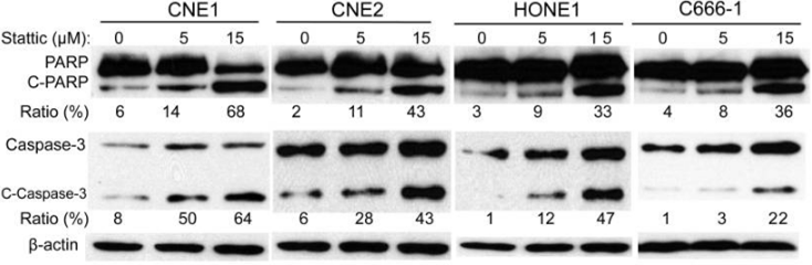
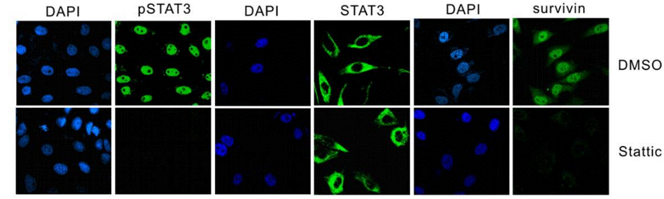
| 実験結果図 | Methods | Biomarkers | 結果図 | PMID |
| Western blot | PARP / C-PARP / Caspase-3 / C-Caspse-3 Survivin / c-Myc / Bcl-xl p-STAT3 / STAT3 |
|
23382914 | |
| Immunofluorescence | p-STAT3 / STAT3 / Survivin |
|
25261365 | |
| Growth inhibition assay | Cell viability |
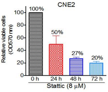
|
23382914 | |
| ELISA | BDNF |
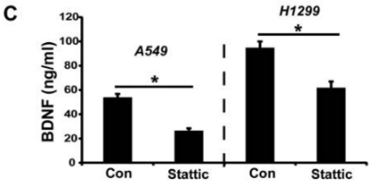
|
27456333 | |
化学情報
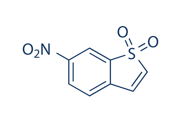
| 分子量 | 211.19 | 化学式 | C8H5NO4S |
| CAS No. | 19983-44-9 | SDF | Download Stattic SDFをダウンロードする |
| Smiles | C1=CC(=CC2=C1C=CS2(=O)=O)[N+](=O)[O-] | ||
| 保管 | |||
|
In vitro |
DMSO : 42 mg/mL ( (198.87 mM); 吸湿したDMSOは溶解度を減少させます。新しいDMSOをご使用ください。) Water : Insoluble Ethanol : Insoluble |
モル濃度計算器 |
|
in vivo Add solvents to the product individually and in order. |
投与溶液組成計算機 | ||||
実験計算
投与溶液組成計算機(クリア溶液)
ステップ1:実験データを入力してください。(実験操作によるロスを考慮し、動物数を1匹分多くして計算・調製することを推奨します)
mg/kg
g
μL
匹
ステップ2:投与溶媒の組成を入力してください。(ロット毎に適した溶解組成が異なる場合があります。詳細については弊社までお問い合わせください)
% DMSO
%
% Tween 80
% ddH2O
%DMSO
%
計算結果:
投与溶媒濃度: mg/ml;
DMSOストック溶液調製方法: mg 試薬を μL DMSOに溶解する(濃度 mg/mL, 注:濃度が当該ロットのDMSO溶解度を超える場合はご連絡ください。 )
投与溶媒調製方法:Take μL DMSOストック溶液に μL PEG300,を加え、完全溶解後μL Tween 80,を加えて完全溶解させた後 μL ddH2O,を加え完全に溶解させます。
投与溶媒調製方法:μL DMSOストック溶液に μL Corn oil,を加え、完全溶解。
注意:1.ストック溶液に沈殿、混濁などがないことをご確認ください;
2.順番通りに溶剤を加えてください。次のステップに進む前に溶液に沈殿、混濁などがないことを確認してから加えてください。ボルテックス、ソニケーション、水浴加熱など物理的な方法で溶解を早めることは可能です。
技術サポート
ストックの作り方、阻害剤の保管方法、細胞実験や動物実験の際に注意すべき点など、製品を取扱う時に問い合わせが多かった質問に対しては取扱説明書でお答えしています。
他に質問がある場合は、お気軽にお問い合わせください。
* 必須
Tags: Statticを買う | Stattic ic50 | Stattic供給者 | Statticを購入する | Stattic費用 | Stattic生産者 | オーダーStattic | Stattic化学構造 | Stattic分子量 | Stattic代理店
納期 国内在庫品:受注日の翌日(15時までの受注分) *北海道、九州、沖縄への配送は受注日より2日以上 を要する場合あり 海外在庫品:受注後1〜2週間