
- 阻害剤
- 研究分野別
- PI3K/Akt/mTOR
- Epigenetics
- Methylation
- Immunology & Inflammation
- Protein Tyrosine Kinase
- Angiogenesis
- Apoptosis
- Autophagy
- ER stress & UPR
- JAK/STAT
- MAPK
- Cytoskeletal Signaling
- Cell Cycle
- TGF-beta/Smad
- 化合物ライブラリー
- 抗体
- 新製品
- お問い合わせ
TPCA-1
別名:GW683965
TPCA-1 (GW683965) is an inhibitor of IKK-2 with IC50 of 17.9 nM in a cell-free assay, inhibits NF-κB pathway, exhibits 22-fold selectivity over IKK-1. TPCA-1 is also an inhibitor of STAT3 and enhances apoptosis.
CAS No. 507475-17-4
文献中Selleckの製品使用例(61)
製品安全説明書
現在のバッチを見る:
純度:
99.85%
99.85
TPCA-1関連製品
シグナル伝達経路
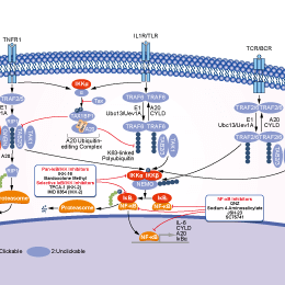
IκB/IKK阻害剤の選択性比較
Cell Data
| Cell Lines | Assay Type | Concentration | Incubation Time | 活性情報 | PMID |
|---|---|---|---|---|---|
| C57BL/6 mouse BMDM cells | Function assay | 0.5 μM | 1 h | Antiinflammatory activity in C57BL/6 mouse BMDM cells assessed as inhibition of LPS-stimulated TNFalpha production at 0.5 uM pretreated for 1 hr before LPS challenge after 8 to 24 hrs by immunoassay | 22533790 |
| C57BL/6 mouse BMDM cells | Cytotoxicity assay | 24 h | Cytotoxicity against C57BL/6 mouse BMDM cells assessed as LDH release after 24 hrs | 22533790 | |
| MDA-MB-231 | Cytotoxicity assay | 3 days | Cytotoxicity against human MDA-MB-231 cells assessed as cell growth inhibition after 3 days by presto blue dye based plate reader method | 27077228 | |
| TC32 | qHTS assay | qHTS of pediatric cancer cell lines to identify multiple opportunities for drug repurposing: Primary screen for TC32 cells | 29435139 | ||
| DAOY | qHTS assay | qHTS of pediatric cancer cell lines to identify multiple opportunities for drug repurposing: Primary screen for DAOY cells | 29435139 | ||
| SJ-GBM2 | qHTS assay | qHTS of pediatric cancer cell lines to identify multiple opportunities for drug repurposing: Primary screen for SJ-GBM2 cells | 29435139 | ||
| A673 | qHTS assay | qHTS of pediatric cancer cell lines to identify multiple opportunities for drug repurposing: Primary screen for A673 cells | 29435139 | ||
| SK-N-MC | qHTS assay | qHTS of pediatric cancer cell lines to identify multiple opportunities for drug repurposing: Primary screen for SK-N-MC cells | 29435139 | ||
| BT-37 | qHTS assay | qHTS of pediatric cancer cell lines to identify multiple opportunities for drug repurposing: Primary screen for BT-37 cells | 29435139 | ||
| NB-EBc1 | qHTS assay | qHTS of pediatric cancer cell lines to identify multiple opportunities for drug repurposing: Primary screen for NB-EBc1 cells | 29435139 | ||
| U-2 OS | qHTS assay | qHTS of pediatric cancer cell lines to identify multiple opportunities for drug repurposing: Primary screen for U-2 OS cells | 29435139 | ||
| Saos-2 | qHTS assay | qHTS of pediatric cancer cell lines to identify multiple opportunities for drug repurposing: Primary screen for Saos-2 cells | 29435139 | ||
| SK-N-SH | qHTS assay | qHTS of pediatric cancer cell lines to identify multiple opportunities for drug repurposing: Primary screen for SK-N-SH cells | 29435139 | ||
| NB1643 | qHTS assay | qHTS of pediatric cancer cell lines to identify multiple opportunities for drug repurposing: Primary screen for NB1643 cells | 29435139 | ||
| LAN-5 | qHTS assay | qHTS of pediatric cancer cell lines to identify multiple opportunities for drug repurposing: Primary screen for LAN-5 cells | 29435139 | ||
| Rh18 | qHTS assay | qHTS of pediatric cancer cell lines to identify multiple opportunities for drug repurposing: Primary screen for Rh18 cells | 29435139 | ||
| OHS-50 | qHTS assay | qHTS of pediatric cancer cell lines to identify multiple opportunities for drug repurposing: Primary screen for OHS-50 cells | 29435139 | ||
| RD | qHTS assay | qHTS of pediatric cancer cell lines to identify multiple opportunities for drug repurposing: Primary screen for RD cells | 29435139 | ||
| MG 63 (6-TG R) | qHTS assay | qHTS of pediatric cancer cell lines to identify multiple opportunities for drug repurposing: Primary screen for MG 63 (6-TG R) cells | 29435139 | ||
| Rh30 | qHTS assay | qHTS of pediatric cancer cell lines to identify multiple opportunities for drug repurposing: Primary screen for Rh30 cells | 29435139 | ||
| Rh41 | qHTS assay | qHTS of pediatric cancer cell lines to identify multiple opportunities for drug repurposing: Primary screen for Rh41 cells | 29435139 | ||
| A673 | qHTS assay | qHTS of pediatric cancer cell lines to identify multiple opportunities for drug repurposing: Confirmatory screen for A673 cells) | 29435139 | ||
| SK-N-MC | qHTS assay | qHTS of pediatric cancer cell lines to identify multiple opportunities for drug repurposing: Confirmatory screen for SK-N-MC cells | 29435139 | ||
| TC32 | qHTS assay | qHTS of pediatric cancer cell lines to identify multiple opportunities for drug repurposing: Confirmatory screen for TC32 cells | 29435139 | ||
| MG 63 (6-TG R) | qHTS assay | qHTS of pediatric cancer cell lines to identify multiple opportunities for drug repurposing: Confirmatory screen for MG 63 (6-TG R) cells | 29435139 | ||
| U-2 OS | qHTS assay | qHTS of pediatric cancer cell lines to identify multiple opportunities for drug repurposing: Confirmatory screen for U-2 OS cells | 29435139 | ||
| SK-N-SH | qHTS assay | qHTS of pediatric cancer cell lines to identify multiple opportunities for drug repurposing: Confirmatory screen for SK-N-SH cells | 29435139 | ||
| 他の多くの細胞株試験データをご覧になる場合はこちらをクリックして下さい | |||||
生物活性
| 製品説明 | TPCA-1 (GW683965) is an inhibitor of IKK-2 with IC50 of 17.9 nM in a cell-free assay, inhibits NF-κB pathway, exhibits 22-fold selectivity over IKK-1. TPCA-1 is also an inhibitor of STAT3 and enhances apoptosis. | ||||||
|---|---|---|---|---|---|---|---|
| Targets |
|
| In Vitro | ||||
| In vitro |
In a time-resolved fluorescence resonance energy transfer assay, TPCA-1 inhibits human IKK-2 activity with an IC50 of 17.9 nM. In addition, TPCA-1 is demonstrated to be ATP-competitive. Besides, TPCA-1 exhibits IC50 values of 400 nM and 3600 nM against IKK-1 and JNK3, respectively. TPCA-1 inhibits the production of TNF-α, IL-6, and IL-8 in a concentration-dependent manner, exhibiting IC50 values of 170, 290, and 320 nM, respectively. [1] TPCA-1 inhibits glioma cell proliferation, as well as TNF-induced RelA (p65) nuclear translocation and NFκB-dependent IL8 gene expression. Importantly, TPCA-1 inhibits IFN-induced gene expression, completely suppressing MX1 and GBP1 gene expression, while having only a minor effect on ISG15 expression. [2] |
|||
|---|---|---|---|---|
| Kinase Assay | IKK-2 Assay | |||
| Recombinant human IKK-2 (residues 1-756) is expressed in baculovirus as an N-terminal GST-tagged fusion protein, and its activity is assessed using a time-resolved fluorescence resonance energy transfer assay. In brief, IKK-2 (5 nM final) diluted in assay buffer (50 mM HEPES, 10 mM MgCl2, 1 mM CHAPS, pH 7.4, with 1 mM DTT and 0.01% w/v BSA) is added to wells containing various concentrations of compound or dimethyl sulfoxide (DMSO) vehicle (3% final). The reaction is initiated by the addition of GST-IκBα substrate (25 nM final)/ATP (1 μM final), in a total volume of 30 μL. The reaction is incubated for 30 min at room temperature, then terminated by the addition of 15 μL of 50 mM EDTA. Detection reagent (15 μL) in buffer (100 mM HEPES, pH 7.4, 150 mM NaCl, and 0.1% w/v BSA) containing antiphosphoserine- IκBα-32/36 monoclonal antibody 12C2, labeled with W-1024 europium chelate, and an allophycocyanin-labeled anti-GST antibody is added, and the reaction is further incubated for 60 min at room temperature. The degree of phosphorylation of GST- IκBαis measured as a ratio of specific 665-nm energy transfer signal to reference europium 620-nm signal, using a Packard Discovery plate reader. | ||||
| 細胞実験 | 細胞株 | U87, MT330, SJ-G2, and GBM6 human glioma lines | ||
| 濃度 | 0-50 μM | |||
| 反応時間 | 3 days | |||
| 実験の流れ | Ten microliters of 3-(4,5-Dimethylthiazol-2-yl)-2,5-diphenyltetrazolium bromide (MTT) from stock solution (10 mg/mL) is added to each well of 96-well plates containing glioma cells and incubated at 37 °C for 2–4 h. Oxidized MTT is solubilized by adding 100 μL of 10% sodium dodecyl sulfate (SDS) in 0.01 N HCL, and plates are incubated at 37 °C for 4 h in a humidified chamber. Plates are read at 570 nm on a plate reader. |
|||
| In Vivo | ||
| In Vivo |
Prophylactic administration of TPCA-1 at 3, 10, or 20 mg/kg, i.p., b.i.d., results in a dose-dependent reduction in the severity of murine collagen-induced arthritis (CIA). The significantly reduced disease severity and delay of disease onset resulting from administration of TPCA-1 at 10 mg/kg, i.p., b.i.d. are comparable to the effects of the antirheumatic drug, etanercept, when administered prophylactically at 4 mg/kg, i.p., every other day. Nuclear localization of p65, as well as levels of IL-1beta, IL-6, TNF-alpha, and interferon-gamma, is significantly reduced in the paw tissue of TPCA-1- and etanercept-treated mice. In addition, administration of TPCA-1 in vivo results in significantly decreased collagen-induced T cell proliferation ex vivo. Therapeutic administration of TPCA-1 at 20 mg/kg, but not at 3 or 10 mg/kg, i.p., b.i.d., significantly reduces the severity of CIA, as does etanercept administration at 12.5 mg/kg, i.p., every other day. [1] |
|
|---|---|---|
| 動物実験 | 動物モデル | Murine collagen-induced arthritis |
| 投与量 | 3, 10, or 20 mg/kg | |
| 投与経路 | Administered via i.p. or b.i.d. | |
化学情報
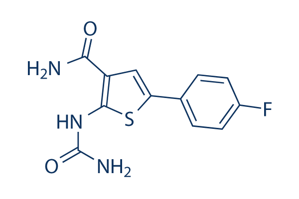
| 分子量 | 279.29 | 化学式 | C12H10FN3O2S |
| CAS No. | 507475-17-4 | SDF | Download TPCA-1 SDFをダウンロードする |
| Smiles | C1=CC(=CC=C1C2=CC(=C(S2)NC(=O)N)C(=O)N)F | ||
| 保管 | |||
|
In vitro |
DMSO : 56 mg/mL ( (200.5 mM); 吸湿したDMSOは溶解度を減少させます。新しいDMSOをご使用ください。) Water : Insoluble Ethanol : Insoluble |
モル濃度計算器 |
|
in vivo Add solvents to the product individually and in order. |
投与溶液組成計算機 | ||||
実験計算
投与溶液組成計算機(クリア溶液)
ステップ1:実験データを入力してください。(実験操作によるロスを考慮し、動物数を1匹分多くして計算・調製することを推奨します)
mg/kg
g
μL
匹
ステップ2:投与溶媒の組成を入力してください。(ロット毎に適した溶解組成が異なる場合があります。詳細については弊社までお問い合わせください)
% DMSO
%
% Tween 80
% ddH2O
%DMSO
%
計算結果:
投与溶媒濃度: mg/ml;
DMSOストック溶液調製方法: mg 試薬を μL DMSOに溶解する(濃度 mg/mL, 注:濃度が当該ロットのDMSO溶解度を超える場合はご連絡ください。 )
投与溶媒調製方法:Take μL DMSOストック溶液に μL PEG300,を加え、完全溶解後μL Tween 80,を加えて完全溶解させた後 μL ddH2O,を加え完全に溶解させます。
投与溶媒調製方法:μL DMSOストック溶液に μL Corn oil,を加え、完全溶解。
注意:1.ストック溶液に沈殿、混濁などがないことをご確認ください;
2.順番通りに溶剤を加えてください。次のステップに進む前に溶液に沈殿、混濁などがないことを確認してから加えてください。ボルテックス、ソニケーション、水浴加熱など物理的な方法で溶解を早めることは可能です。
技術サポート
ストックの作り方、阻害剤の保管方法、細胞実験や動物実験の際に注意すべき点など、製品を取扱う時に問い合わせが多かった質問に対しては取扱説明書でお答えしています。
他に質問がある場合は、お気軽にお問い合わせください。
* 必須
Tags: TPCA-1を買う | TPCA-1 ic50 | TPCA-1供給者 | TPCA-1を購入する | TPCA-1費用 | TPCA-1生産者 | オーダーTPCA-1 | TPCA-1化学構造 | TPCA-1分子量 | TPCA-1代理店
納期 国内在庫品:受注日の翌日(15時までの受注分) *北海道、九州、沖縄への配送は受注日より2日以上 を要する場合あり 海外在庫品:受注後1〜2週間