
- 阻害剤
- 研究分野別
- PI3K/Akt/mTOR
- Epigenetics
- Methylation
- Immunology & Inflammation
- Protein Tyrosine Kinase
- Angiogenesis
- Apoptosis
- Autophagy
- ER stress & UPR
- JAK/STAT
- MAPK
- Cytoskeletal Signaling
- Cell Cycle
- TGF-beta/Smad
- 化合物ライブラリー
- 抗体
- 新製品
- お問い合わせ
Tubastatin A HCl
Tubastatin A HCl is a potent and selective HDAC6 inhibitor with IC50 of 15 nM in a cell-free assay. It is selective (1000-fold more) against all other isozymes except HDAC8 (57-fold more).
CAS No. 1310693-92-5
文献中Selleckの製品使用例(47)
製品安全説明書
現在のバッチを見る:
純度:
99.30%
99.30
Tubastatin A HCl関連製品
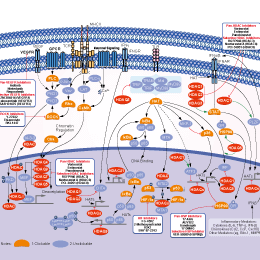
シグナル伝達経路
HDAC阻害剤の選択性比較
Cell Data
| Cell Lines | Assay Type | Concentration | Incubation Time | 活性情報 | PMID |
|---|---|---|---|---|---|
| neuron cultures | Kinase assay | 2.5 μM | induces α-tubulin hyperacetylation | 20614936 | |
| neuron cultures | Function assay | ~10 μM | protects against glutathione depletion-induced oxidative stress | 20614936 | |
| 134/04 | Function assay | 7.5 µM | impairs myotube formation | 22174839 | |
| C2C12 | Function assay | 7.5 µM | impairs myotube formation | 22174839 | |
| HaCaT keratinocytes | Function assay | 10 μM | blocks arsenite from inducing Nrf2 protein translation | 22367689 | |
| JURL-MK1 | Function assay | 10 μM | enhances cell adhesivity to fibronectin | 23022583 | |
| CML-T1 | Function assay | 10 μM | enhances cell adhesivity to fibronectin | 23022583 | |
| K562 | Function assay | 10 μM | enhances cell adhesivity to fibronectin | 23022583 | |
| HL-60 | Function assay | 10 μM | enhances cell adhesivity to fibronectin | 23022583 | |
| KMCH | Growth inhibitory assay | ~10 μM | decreases proliferation and anchorage-independent growth | 23370327 | |
| THP-1 | Function assay | ~10 μM | inhibits TNF-α and IL-6 secretion | 23541634 | |
| RAW 264.7 | Function assay | ~10 μM | attenuates NO production | 23541634 | |
| HT3 | Function assay | ~5 μM | induces the differential α-tubulin acetylation | 23698468 | |
| SiHa | Function assay | ~5 μM | induces the differential α-tubulin acetylation | 23698468 | |
| CaSki | Function assay | ~5 μM | induces the differential α-tubulin acetylation | 23698468 | |
| SiHa | Function assay | ~5 μM | inhibits Thapsigargin- or EGF-induced SOCE activation | 23698468 | |
| CaSki | Function assay | ~5 μM | inhibits Thapsigargin- or EGF-induced SOCE activation | 23698468 | |
| MCF-7 | Growth inhibitory assay | 30 μM | IC50=15 μM | 23798680 | |
| MCF-7 | Function assay | 30 μM | increases the microtubule acetylation level. | 23798680 | |
| MCF-7 | Function assay | 30 μM | stabilizes microtubules against cold-induced depolymerization | 23798680 | |
| MCF-7 | Function assay | 15 μM | stabilizes microtubules against nocodazole-induced disassembly | 23798680 | |
| MCF-7 | Function assay | 30 μM | alteres the assembly dynamics of interphase microtubules | 23798680 | |
| MCF-7 | Function assay | 30 μM | increases the binding of HDAC6 with interphase microtubules | 23798680 | |
| PC12 | Function assay | ~3 μM | up-regulates anti-oxidative gene expression related to transcription factor XBP1s | 24909686 | |
| PC12 | Growth inhibitory assay | ~3 μM | reverse H2O2-induced growth inhibition | 24909686 | |
| HEK293T | Function assay | ~3 μM | up-regulated XBP1s protein level | 24909686 | |
| HEK293T | Function assay | ~3 μM | delays XBP1s protein degradation via acetylation-mediated proteasomal degradation | 24909686 | |
| Huh7 | Function assay | ~5 μM | suppresses proliferation of hepatitis C virus replicon with EC50 = 0.3 μM | 25108326 | |
| SKMEL21 | Growth inhibitory assay | ~500 nM | inhibits cell proliferation | 25957812 | |
| SKMEL103 | Growth inhibitory assay | ~500 nM | inhibits cell proliferation | 25957812 | |
| SKMEL28 | Growth inhibitory assay | ~500 nM | inhibits cell proliferation | 25957812 | |
| WM164 | Growth inhibitory assay | ~500 nM | inhibits cell proliferation | 25957812 | |
| WM1361a | Growth inhibitory assay | ~500 nM | inhibits cell proliferation | 25957812 | |
| WM1366 | Growth inhibitory assay | ~500 nM | inhibits cell proliferation | 25957812 | |
| WM793 | Growth inhibitory assay | ~500 nM | inhibits cell proliferation | 25957812 | |
| WM35 | Growth inhibitory assay | ~500 nM | inhibits cell proliferation | 25957812 | |
| WM983a | Growth inhibitory assay | ~500 nM | inhibits cell proliferation | 25957812 | |
| WM793 | Function assay | ~6 μM | induce G1 arrest | 25957812 | |
| WM164 | Function assay | ~6 μM | induce G1 arrest | 25957812 | |
| WM983a | Function assay | ~6 μM | induce G1 arrest | 25957812 | |
| WM164 | Function assay | ~3 μM | augments expression of MHC class I and melanoma associated antigens | 25957812 | |
| WM983a | Function assay | ~3 μM | augments expression of MHC class I and melanoma associated antigens | 25957812 | |
| IPC298 | Function assay | ~3 μM | augments expression of MHC class I and melanoma associated antigens | 25957812 | |
| SKMEL30 | Function assay | ~3 μM | augments expression of MHC class I and melanoma associated antigens | 25957812 | |
| 293T | Function assay | ~2 μg/ml | induces PTEN expression and membrane translocation | 26279303 | |
| SACC-83 | Function assay | ~2 μg/ml | induces PTEN expression and membrane translocation | 26279303 | |
| 293T | Function assay | ~2 μg/ml | induces PTEN acetylation at K163 | 26279303 | |
| U-87 MG | Function assay | ~2 μg/ml | inhibits the migration and invasion | 26279303 | |
| U-87 MG | Function assay | ~10 μM | inhibits AKT phosphorylation | 26279303 | |
| U-87 MG | Growth inhibitory assay | ~10 μM | inhibits cell growth | 26279303 | |
| TCa83 | Function assay | induces PTEN expression and membrane translocation | 26279303 | ||
| 他の多くの細胞株試験データをご覧になる場合はこちらをクリックして下さい | |||||
生物活性
| 製品説明 | Tubastatin A HCl is a potent and selective HDAC6 inhibitor with IC50 of 15 nM in a cell-free assay. It is selective (1000-fold more) against all other isozymes except HDAC8 (57-fold more). | ||||
|---|---|---|---|---|---|
| Targets |
|
| In Vitro | ||||
| In vitro | Tubastatin A is substantially selective for all 11 HDAC isoforms and maintains over 1000-fold selectivity against all isoforms excluding HDAC8, where it has approximately 57-fold selectivity. In homocysteic acid (HCA) induced neurodegeneration assays, Tubastatin A displays dose-dependent protection against HCA-induced neuronal cell death starting at 5 μM with near complete protection at 10 μM. [1] At 100 ng/mL Tubastatin A increases Foxp3+ T-regulatory cells (Tregs) suppression of T cell proliferation in vitro. [2] Tubastatin A treatment in C2C12 cells would lead to myotube formation impairment when alpha-tubulin is hyperacetylated early in the myogenic process; however, myotube elongation occurs when alpha-tubulin is hyeperacetylated in myotubes. [3] A recent study indicates that Tubastatin A treatment increases cell elasticity as revealed by atomic force microscopy (AFM) tests without exerting drastic changes to the actin microfilament or microtubule networks in mouse ovarian cancer cell lines, MOSE-E and MOSE-L. [4] | |||
|---|---|---|---|---|
| Kinase Assay | Enzyme Inhibition Assays | |||
| Enzyme inhibition assays are performed by the Reaction Biology Corporation, Malvern, PA, using the Reaction Biology HDAC Spectrum platform. (www.reactionbiology.com) The HDAC1, 2, 4, 5, 6, 7, 8, 9, 10, and 11 assays use isolated recombinant human protein; HDAC3/NcoR2 complex is used for the HDAC3 assay. Substrate for HDAC1, 2, 3, 6, 10, and 11 assays is a fluorogenic peptide from p53 residues 379-382 (RHKKAc); substrate for HDAC8 is fluorogenic diacyl peptide based on residues 379-382 of p53 (RHKAcKAc). Acetyl-Lys (trifluoroacetyl)-AMC substrate is used for HDAC4, 5, 7, and 9 assays. Tubastatin A is dissolved in DMSO and tested in 10-dose IC50 mode with 3-fold serial dilution starting at 30 μM. Control Compound Trichostatin A (TSA) is tested in a 10-dose IC50 with 3-fold serial dilution starting at 5 μM. IC50 values are extracted by curve-fitting the dose/response slopes. | ||||
| 細胞実験 | 細胞株 | Primary cortical neuron of fetal Sprague-Dawley rats (embryonic day 17) | ||
| 濃度 | 0-10 μM | |||
| 反応時間 | 24 hours | |||
| 実験の流れ | Primary cortical neuron cultures are obtained from the cerebral cortex of fetal Sprague-Dawley rats (embryonic day 17) as described previously. All experiments are initiated 24 hours after plating. Under these conditions, the cells are not susceptible to glutamate-mediated excitotoxicity. For cytotoxicity studies, cells are rinsed with warm PBS and then placed in minimum essential medium (Invitrogen) containing 5.5 g/L glucose, 10% fetal calf serum, 2 mM L-glutamine, and 100 μM cystine. Oxidative stress is induced by the addition of the glutamate analogue homocysteate (HCA; 5 mM) to the media. HCA is diluted from 100-fold concentrated solutions that are adjusted to pH 7.5. In combination with HCA, neurons are treated with Tubastatin A at the indicated concentrations. Viability is assessed after 24 hours by MTT assay (3-(4,5-dimethylthiazol-2-yl)-2,5-diphenyltetrazolium bromide) method. | |||
| 実験結果図 | Methods | Biomarkers | 結果図 | PMID |
| Western blot | EGFR / p-AKT / AKT / p-ERK / ERK |
|
29665050 | |
| Immunofluorescence | α-tubulin / Acetylated tubulin HDAC6 |
|
23798680 | |
| In Vivo | ||
| In Vivo | Daily treatment of Tubastatin A at 0.5mg/kg inhibits HDAC6 to promote Tregs suppressive activity in mouse models of inflammation and autoimmunity, including multiple forms of experimental colitis and fully major histocompatibility complex (MHC)-incompatible cardiac allograft rejection. [2] | |
|---|---|---|
| 動物実験 | 動物モデル | Na?ve CD45RBhi CD4+ CD25- cells (1 × 10⁶) from WT or HDAC6-/- mice Are injected i.p. into B6/Rag1-/-mice. |
| 投与量 | 0.5 mg/kg | |
| 投与経路 | Tubastatin A is injected i.p. daily. | |
化学情報
| 分子量 | 371.86 | 化学式 | C20H21N3O2.HCl |
| CAS No. | 1310693-92-5 | SDF | Download Tubastatin A HCl SDFをダウンロードする |
| Smiles | CN1CCC2=C(C1)C3=CC=CC=C3N2CC4=CC=C(C=C4)C(=O)NO.Cl | ||
| 保管 | |||
|
In vitro |
DMSO : 100 mg/mL ( (268.91 mM); 吸湿したDMSOは溶解度を減少させます。新しいDMSOをご使用ください。) Water : Insoluble Ethanol : Insoluble |
モル濃度計算器 |
|
in vivo Add solvents to the product individually and in order. |
投与溶液組成計算機 | ||||
実験計算
投与溶液組成計算機(クリア溶液)
ステップ1:実験データを入力してください。(実験操作によるロスを考慮し、動物数を1匹分多くして計算・調製することを推奨します)
mg/kg
g
μL
匹
ステップ2:投与溶媒の組成を入力してください。(ロット毎に適した溶解組成が異なる場合があります。詳細については弊社までお問い合わせください)
% DMSO
%
% Tween 80
% ddH2O
%DMSO
%
計算結果:
投与溶媒濃度: mg/ml;
DMSOストック溶液調製方法: mg 試薬を μL DMSOに溶解する(濃度 mg/mL, 注:濃度が当該ロットのDMSO溶解度を超える場合はご連絡ください。 )
投与溶媒調製方法:Take μL DMSOストック溶液に μL PEG300,を加え、完全溶解後μL Tween 80,を加えて完全溶解させた後 μL ddH2O,を加え完全に溶解させます。
投与溶媒調製方法:μL DMSOストック溶液に μL Corn oil,を加え、完全溶解。
注意:1.ストック溶液に沈殿、混濁などがないことをご確認ください;
2.順番通りに溶剤を加えてください。次のステップに進む前に溶液に沈殿、混濁などがないことを確認してから加えてください。ボルテックス、ソニケーション、水浴加熱など物理的な方法で溶解を早めることは可能です。
技術サポート
ストックの作り方、阻害剤の保管方法、細胞実験や動物実験の際に注意すべき点など、製品を取扱う時に問い合わせが多かった質問に対しては取扱説明書でお答えしています。
他に質問がある場合は、お気軽にお問い合わせください。
* 必須
Tags: Tubastatin A HClを買う | Tubastatin A HCl ic50 | Tubastatin A HCl供給者 | Tubastatin A HClを購入する | Tubastatin A HCl費用 | Tubastatin A HCl生産者 | オーダーTubastatin A HCl | Tubastatin A HCl化学構造 | Tubastatin A HCl分子量 | Tubastatin A HCl代理店
納期 国内在庫品:受注日の翌日(15時までの受注分) *北海道、九州、沖縄への配送は受注日より2日以上 を要する場合あり 海外在庫品:受注後1〜2週間