|
受注:045-509-1970 |
技術サポート:tech@selleck.co.jp 平日9:00〜18:00 1営業日以内にご連絡を差し上げます |
生物学的記述
| Specificity | Tenascin C Antibody [L15L8] detects endogenous levels of total Tenascin C protein. |
|---|---|
| Background | Tenascin C is a large, multimodular, hexabrachion‑type extracellular matrix glycoprotein and founding member of the tenascin family, characterized by a central coiled‑coil assembly domain that mediates oligomerization, a series of epidermal growth factor–like repeats, a variable number of alternatively spliced fibronectin type III (FNIII) domains, and a C‑terminal fibrinogen‑like globe that together create multiple interaction surfaces for matrix components, growth factors, and cell‑surface receptors. Restricted expression during embryogenesis and virtual absence from most healthy adult tissues contrast with strong re‑induction at sites of tissue remodeling, chronic inflammation, and tumor stroma, where tenascin C functions as an adhesion‑modulating matrix organizer that weakens fibronectin‑mediated adhesion, destabilizes actin stress fibers, and promotes cell rounding, proliferation, and motility to support processes such as epithelial–mesenchymal transition, wound repair, and invasive growth. The protein interacts with fibronectin in the solid phase and with integrins including αvβ3, α9β1, and α2β1, as well as cell‑surface heparan sulfate proteoglycans, and distinct FNIII repeats and splice variants differentially engage these receptors to activate or repress downstream signaling pathways such as focal adhesion kinase (FAK), Src, Rho GTPases, and ERK/MAPK, enabling context‑dependent control of migration and proliferation. In glioblastoma models, adhesion of tumor cells to tenascin C via αvβ3 on a fibronectin substratum inhibits stable spreading yet enhances growth by down‑regulating tropomyosin‑1, destabilizing actin filaments, up‑regulating endothelin receptor type A and activating endothelin‑driven ERK1/2 and c‑Fos, and relieving Wnt pathway inhibition through repression of Dickkopf‑1, which stabilizes β‑catenin and increases expression of the β‑catenin target Id2, correlating tenascin C and Id2 expression with high astrocytoma malignancy. More broadly, tenascin C modulates growth factor and cytokine availability and signaling by binding to and organizing matrix‑associated ligands, by inducing pro‑inflammatory mediators, and by functioning as an endogenous danger‑associated molecular pattern that activates Toll‑like receptor 4 and drives AKT/PI3K‑ and NF‑κB‑dependent production of inflammatory cytokines, thereby establishing an autocrine loop that sustains chronic inflammation in autoimmune disease and supports tumor invasion and egress. In tissue injury and cardiovascular remodeling, tenascin C expression rises in myocardium, vessel walls, and interstitial stroma exposed to altered mechanical load, where it influences mechanotransduction, cell–matrix resilience, and fibroblast proliferation, integrating cues from TGF‑β, angiotensin II, and mechanical stretch to coordinate fibrotic and regenerative responses. Within the central nervous system, tenascin C is produced by radial glia, astrocytes, and oligodendrocyte precursor cells during development, where it shapes astrocyte lineage progression by regulating patterning genes such as Nkx6.1 and Nkx2.2 and their downstream effector sulfatase‑1, and it persists or reappears in adult neurogenic niches, gliomas, and injury sites, where it guides axon growth, modulates synaptic plasticity, and contributes to glial scar formation and neuroinflammation. |
使用情報
| Application | WB, IP, IF | Dilution |
|
||||||
|---|---|---|---|---|---|---|---|---|---|
| Reactivity | Human | ||||||||
| Source | Rabbit Monoclonal Antibody | MW | 241 kDa | ||||||
| Storage Buffer | PBS, pH 7.2+50% Glycerol+0.05% BSA+0.01% NaN3 | Storage (from the date of receipt) |
-20°C (avoid freeze-thaw cycles), 2 years | ||||||
References
|
Application Data
WB
Validated by Selleck
-
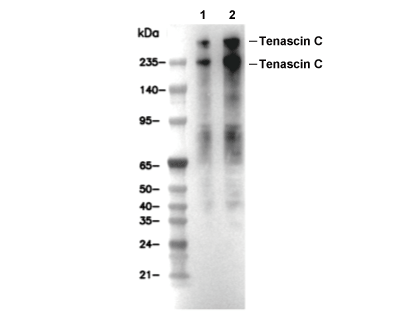 Lane 1: U87MG, Lane 2: U87MG (BFA, 5ug/mL, 6 h)