|
受注:045-509-1970 |
技術サポート:tech@selleck.co.jp 平日9:00〜18:00 1営業日以内にご連絡を差し上げます |
生物学的記述
| Specificity | TLR4 Antibody [L6M8] detects endogenous levels of total TLR4 protein. |
|---|---|
| Background | TLR4 (Toll‑like receptor 4) is a type I transmembrane pattern‑recognition receptor of the Toll/IL‑1 receptor (TIR) family that detects Gram‑negative bacterial lipopolysaccharide and selected endogenous damage‑associated ligands and initiates innate immune signaling programs that couple microbial sensing to inflammatory and antiviral gene expression. The receptor contains an N‑terminal extracellular leucine‑rich repeat ectodomain that forms a curved solenoid scaffold for ligand recognition with the accessory protein MD‑2, a single transmembrane helix, and a cytoplasmic TIR domain that nucleates adaptor recruitment and signal propagation; functional signaling requires assembly of an LPS–CD14–MD‑2–TLR4 complex and dimerization of two TLR4–MD‑2 units at the plasma membrane. TLR4 expression is prominent on myeloid cells such as macrophages and dendritic cells and is also present on endothelial and epithelial cells where it operates as a primary sensor of circulating endotoxin and of endogenous alarmins, driving rapid transcriptional responses that include TNF, IL‑6, IL‑1 family cytokines, chemokines, co‑stimulatory molecules, and interferon‑regulated genes. Ligand engagement first activates a MyD88‑dependent cascade at the plasma membrane through recruitment of the TIR adaptors TIRAP/MAL and MyD88 to the TLR4 TIR domain, followed by assembly of IRAK kinases and TRAF6, activation of TAK1, and stimulation of the IKK complex and MAPKs, which leads to IκB degradation and NF‑κB nuclear translocation together with AP‑1 activation and robust production of pro‑inflammatory mediators. Subsequent endocytosis of the TLR4 complex and signaling from early endosomes uses TRAM and TRIF as TIR adaptors to activate TBK1 and IKKε, phosphorylate IRF3, and induce type I interferon and interferon‑inducible genes, while also engaging a TRIF–RIP1–IKK axis that reinforces NF‑κB‑ and MAPK‑dependent responses, so that TLR4 couples surface and endosomal platforms to integrate pro‑inflammatory and antiviral outputs. These MyD88‑ and TRIF‑dependent branches are modulated by regulatory proteins such as SIGIRR, SOCS, and A20 and by scaffold proteins including GIV/Girdin, which dampen excessive macrophage activation and shape the magnitude and duration of cytokine release, linking TLR4 signal strength to protection from infection on one side and to tissue‑damaging inflammation on the other. |
使用情報
| Application | WB, IHC, IF, FCM, ChIP, ELISA | Dilution |
|
||||||||
|---|---|---|---|---|---|---|---|---|---|---|---|
| Reactivity | Human, Mouse, Rat, Porcine, Bovine, Mammal | ||||||||||
| Source | Mouse Monoclonal Antibody | MW | 95.7 kDa | ||||||||
| Storage Buffer | PBS, pH 7.2+50% Glycerol+0.05% BSA+0.01% NaN3 | Storage (from the date of receipt) |
-20°C (avoid freeze-thaw cycles), 2 years | ||||||||
References
|
Application Data
WB
Validated by Selleck
-
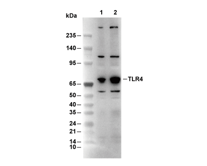 Lane 1: Raw 264.7, Lane 2: K562