|
受注:045-509-1970 |
技術サポート:tech@selleck.co.jp 平日9:00〜18:00 1営業日以内にご連絡を差し上げます |
生物学的記述
| Specificity | Angiomotin Antibody [M16G16] detects endogenous levels of total Angiomotin protein. |
|---|---|
| Background | Angiomotin (AMOT) is a junction‑associated scaffold protein belonging to the Motin family, which includes AMOTL1 and AMOTL2, and is widely expressed in epithelial and endothelial tissues where it coordinates cell–cell adhesion, polarity, and migration. It exists primarily as two isoforms generated by alternative splicing, a full‑length p130 form and a shorter, amino‑terminally truncated p80 variant, both of which share conserved N‑terminal coiled‑coil and C‑terminal PDZ‑domain‑like motifs that mediate interactions with membrane‑associated and cytoskeletal proteins. AMOT directly binds the transcriptional co‑activators YAP and TAZ, sequestering them in the cytoplasm and limiting their nuclear entry and TEAD‑dependent transcription. This interaction can occur independently of canonical Hippo‑pathway phosphorylation. In endothelial cells, AMOT acts downstream of GPCR signaling and is a direct substrate of Lats1/2 kinases. Phosphorylation of conserved motifs in AMOT‑p130 disrupts its association with F‑actin and stress fibers, reduces focal adhesions, impairs migration, and attenuates angiogenesis. AMOT also interfaces with small‑GTPase networks by scaffolding components around junctional complexes, including interactions with Merlin and regulators of Rac1, and modulates actin organization and cytoskeletal dynamics that affect cell shape, collective migration, and barrier integrity. AMOT‑p130 differentially regulates Rho‑ and actin‑dependent cytoskeletal remodeling in epithelial and cancer contexts. In some breast cancer models, AMOT overexpression correlates with increased proliferation, invasion, and metastasis‑linked motility, while in other settings it correlates with reduced YAP‑driven growth. Angiomotin isoforms show differential expression, are subject to post‑translational modification by Lats1/2, and compete with other Motin‑family members. |
使用情報
| Application | WB, IP, IHC, IF, FCM | Dilution |
|
||||||||||
|---|---|---|---|---|---|---|---|---|---|---|---|---|---|
| Reactivity | Human, Mouse, Rat | ||||||||||||
| Source | Rabbit Monoclonal Antibody | MW | 140 kDa, 80 kDa | ||||||||||
| Storage Buffer | PBS, pH 7.2+50% Glycerol+0.05% BSA+0.01% NaN3 | Storage (from the date of receipt) |
-20°C (avoid freeze-thaw cycles), 2 years | ||||||||||
References
|
Application Data
WB
Validated by Selleck
-
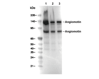 Lane 1: 293T, Lane 2: OVCAR3, Lane 3: C2C12