
|
受注:045-509-1970 |
技術サポート:tech@selleck.co.jp 平日9:00〜18:00 1営業日以内にご連絡を差し上げます |
生物学的記述
| Specificity | Brachyury/Bry Antibody (Rabbit mAb) [K13E20] detects endogenous levels of total Brachyury/Bry protein. |
|---|---|
| Background | Brachyury, encoded by the T gene, is the founder member of the T‑box transcription factor family and a key regulator of posterior mesoderm formation and axial development during vertebrate gastrulation, where its transient expression in primitive streak‑derived nascent and migrating mesoderm cells defines axial and paraxial mesoderm lineages and coordinates morphogenetic movements that generate the notochord and structures caudal to the forelimb. The protein contains a conserved T‑box DNA‑binding domain that adopts a seven‑stranded β‑barrel architecture related to the immunoglobulin fold, with inserted α‑helices that form a composite surface recognizing a highly conserved palindromic consensus sequence predominantly in the minor groove; structural analyses show that Brachyury dimerizes on DNA via contacts between specific β‑strand loops and helices, and that the dimer interface and DNA‑contacting residues are tuned to bind and regulate enhancers of target genes such as fibroblast growth factor 4 (FGF4/eFGF) and Brachyury‑induced homeobox (Bix) genes, placing Brachyury at the head of gene regulatory modules controlling mesoderm differentiation. Genetic studies in mouse demonstrate that homozygous T mutants die in midgestation with accumulation of mesodermal cells at the primitive streak, loss of posterior mesoderm and abnormal notochord, confirming that Brachyury is required for mesoderm specification and for the epithelial‑to‑mesenchymal transitions and migratory behaviors that drive axial extension, while overexpression of Brachyury mRNA is sufficient to induce mesodermal characteristics and upregulate eFGF, linking its transcriptional activity directly to FGF signaling pathways. Charting of Brachyury‑mediated developmental pathways during early mouse embryogenesis using genome‑wide occupancy and expression profiling reveals that Brachyury binds enhancers near genes involved in Wnt, FGF, TGF‑β and BMP signaling and in cytoskeletal dynamics, and that its targets integrate positional information along the anterior–posterior axis with cell movement programs, establishing Brachyury as a central node in the network that couples morphogen gradients to mesoderm fate and germ layer patterning. In humans, T is expressed in the notochord and adult chordoma tumors, and Brachyury protein serves as a highly specific diagnostic marker for notochord‑derived neoplasms; allele association studies identify a common intronic polymorphism (TIVS7‑2) in human T that shows significant transmission disequilibrium with spina bifida, and combined data from British and Dutch families support a modest but reproducible association between this T allele and susceptibility to neural tube defects, implicating variants in Brachyury in multifactorial NTD risk while not acting as a sole causative factor. Beyond development, Brachyury overexpression in a range of human carcinomas correlates with epithelial–mesenchymal transition, tumor metastasis, cancer stem cell marker expression and resistance to chemotherapy and radiotherapy, and mechanistic analyses indicate that Brachyury participates in EMT‑related signaling circuits involving fibroblast growth factor and transforming growth factor‑β, where its transcriptional programs upregulate mesenchymal genes and downregulate epithelial markers to promote invasiveness and spheroid formation. Targeting of Brachyury‑positive tumor cells with recombinant yeast‑Brachyury vector‑based vaccines activates Brachyury‑specific CD4⁺ and CD8⁺ T cells capable of lysing human tumor cells expressing Brachyury, and structural studies of human Brachyury DNA‑recognition complexes have begun to identify small‑molecule ligands that bind its T‑box domain, suggesting emerging routes to therapeutic modulation of Brachyury activity in chordoma and EMT‑driven cancers. Overall, Brachyury is a structurally distinctive T‑box transcription factor whose β‑barrel/helix DNA‑binding domain and dimerization interface confer selective recognition of palindromic T‑box sites, whose developmental role is to specify and drive posterior mesoderm and notochord morphogenesis through FGF‑centered gene networks, whose human polymorphisms influence neural tube defect susceptibility, and whose re‑expression and upregulation in carcinomas mechanistically link embryonic mesoderm programs to EMT, stemness, metastasis and immunotherapeutic targeting. |
使用情報
| Application | WB, IP, IF, FCM | Dilution |
|
||||||||
|---|---|---|---|---|---|---|---|---|---|---|---|
| Reactivity | Human | ||||||||||
| Source | Rabbit Monoclonal Antibody | MW | 47 kDa | ||||||||
| Storage Buffer | PBS, pH 7.2+50% Glycerol+0.05% BSA+0.01% NaN3 | Storage (from the date of receipt) |
-20°C (avoid freeze-thaw cycles), 2 years | ||||||||
| WB |
Experimental Protocol:
Sample preparation
1. Tissue: Lyse the tissue sample by adding an appropriate volume of ice-cold RIPA/Nuclear Lysis Buffer (containing Protease Inhibitor Cocktail),and homogenize the tissue at a low temperature or lyse it by sonication on ice, then incubate on ice for 30 minutes. 2. Adherent cell: Aspirate the culture medium and wash the cells with ice-cold PBS twice. Lyse the cells by adding an appropriate volume of RIPA/Nuclear Lysis Buffer (containing Protease Inhibitor Cocktail) , sonicate to lyse the cells, and incubate on ice for 30 minutes. 3. Suspension cell: Transfer the culture medium to a pre-cooled centrifuge tube. Centrifuge and aspirate the supernatant. Wash the cells with ice-cold PBS twice. Lyse the cells by adding an appropriate volume of RIPA/Nuclear Lysis Buffer (containing Protease Inhibitor Cocktail) , sonicate to lyse the cells, and incubate on ice for 30 minutes. 4. Place the lysate into a pre-cooled microcentrifuge tube. Centrifuge at 4°C for 15 min. Collect the supernatant;
5. Remove a small volume of lysate to determine the protein concentration;
6. Combine the lysate with protein loading buffer. Boil 20 µL sample under 95-100°C for 5 min. Centrifuge for 5 min after cool down on ice.
Electrophoretic separation
1. According to the concentration of extracted protein, load appropriate amount of protein sample and marker onto SDS-PAGE gels for electrophoresis. Recommended separating gel (lower gel) concentration: 10%. Reference Table for Selecting SDS-PAGE Separation Gel Concentrations 2. Power up 80V for 30 minutes. Then the power supply is adjusted (110 V~150 V), the Marker is observed, and the electrophoresis can be stopped when the indicator band of the predyed protein Marker where the protein is located is properly separated. (Note that the current should not be too large when electrophoresis, too large current (more than 150 mA) will cause the temperature to rise, affecting the result of running glue. If high currents cannot be avoided, an ice bath can be used to cool the bath.)
Transfer membrane
1. Take out the converter, soak the clip and consumables in the pre-cooled converter;
2. Activate PVDF membrane with methanol for 1 min and rinse with transfer buffer;
3. Install it in the order of "black edge of clip - sponge - filter paper - filter paper - glue -PVDF membrane - filter paper - filter paper - sponge - white edge of clip"; 4. The protein was electrotransferred to PVDF membrane. ( 0.45 µm PVDF membrane is recommended ) Reference Table for Selecting PVDF Membrane Pore Size Specifications Recommended conditions for wet transfer: 200 mA, 120 min. ( Note that the transfer conditions can be adjusted according to the protein size. For high-molecular-weight proteins, a higher current and longer transfer time are recommended. However, ensure that the transfer tank remains at a low temperature to prevent gel melting.)
Block
1. After electrotransfer, wash the film with TBST at room temperature for 5 minutes;
2. Incubate the film in the blocking solution for 1 hour at room temperature;
3. Wash the film with TBST for 3 times, 5 minutes each time.
Antibody incubation
1. Use primary antibody dilution buffer to prepare the primary antibody working liquid (recommended dilution ratio for primary antibody 1:1000), gently shake and incubate with the film at 4°C overnight; 2. Wash the film with TBST 3 times, 5 minutes each time;
3. Add the secondary antibody to the blocking solution and incubate with the film gently at room temperature for 1 hour;
4. After incubation, wash the film with TBST 3 times for 5 minutes each time.
Antibody staining
1. Add the prepared ECL luminescent substrate (or select other color developing substrate according to the second antibody) and mix evenly;
2. Incubate with the film for 1 minute, remove excess substrate (keep the film moist), wrap with plastic film, and expose in the imaging system.
|
| IF |
Experimental Protocol:
Sample Preparation
1. Adherent Cells: Place a clean, sterile coverslip in a culture dish. Once the cells grow to near confluence as a monolayer, remove the coverslip for further use.
2. Suspension Cells: Seed the cells onto a clean, sterile slide coated with poly-L-lysine.
3. Frozen Sections: Allow the slide to thaw at room temperature. Wash it with pure water or PBS for 2 times, 3 minutes each time.
4. Paraffin Sections: Deparaffinization and rehydration. Wash the slide with pure water or PBS for 3 times, 3 minutes each time. Then perform antigen retrieval.
Fixation
1. Fix the cell coverslips/spots or tissue sections at room temperature using a fixative such as 4% paraformaldehyde (4% PFA) for 10-15 minutes.
2. Wash the sample with PBS for 3 times, 3 minutes each time.
Permeabilization
1.Add a detergent such as 0.1–0.3% Triton X-100 to the sample and incubate at room temperature for 10–20 minutes.
(Note: This step is only required for intracellular antigens. For antigens expressed on the cell membrane, this step is unnecessary.)
Wash the sample with PBS for 3 times, 3 minutes each time.
Blocking
Add blocking solution and incubate at room temperature for at least 1 hour. (Common blocking solutions include: serum from the same source as the secondary antibody, BSA, or goat serum.)
Note: Ensure the sample remains moist during and after the blocking step to prevent drying, which can lead to high background.
Immunofluorescence Staining (Day 1)
1. Remove the blocking solution and add the diluted primary antibody.
2. Incubate the sample in a humidified chamber at 4°C overnight.
Immunofluorescence Staining (Day 2)
1. Remove the primary antibody and wash with PBST for 3 times, 5 minutes each time.
2. Add the diluted fluorescent secondary antibody and incubate in the dark at 4°C for 1–2 hours.
3. Remove the secondary antibody and wash with PBST for 3 times, 5 minutes each time.
4. Add diluted DAPI and incubate at room temperature in the dark for 5–10 minutes.
5. Wash with PBST for 3 times, 5 minutes each time.
Mounting
1. Mount the sample with an anti-fade mounting medium.
2. Allow the slide to dry at room temperature overnight in the dark.
3. Store the slide in a slide storage box at 4°C, protected from light.
|
References
|
Application Data
WB
Validated by Selleck
-
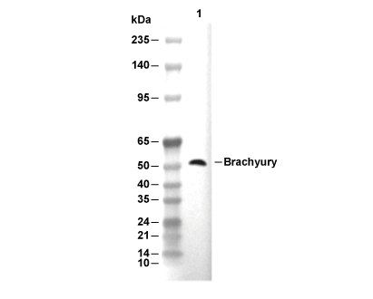 Lane 1: NCI-H460