
|
受注:045-509-1970 |
技術サポート:tech@selleck.co.jp 平日9:00〜18:00 1営業日以内にご連絡を差し上げます |
生物学的記述
| Specificity | BSCL2/Seipin Antibody (Rabbit mAb) [N16E20] detects endogenous levels of total BSCL2/Seipin protein. |
|---|---|
| Background | Seipin, encoded by BSCL2, is a conserved homo‑oligomeric endoplasmic reticulum membrane protein that concentrates at ER–lipid droplet junctions and operates as a central organizer of lipid droplet biogenesis and adipocyte lipid storage, with loss‑of‑function mutations causing the most severe form of congenital generalized lipodystrophy characterized by near‑complete absence of adipose tissue, extreme insulin resistance and dyslipidemia. The protein forms large ring‑like oligomers of defined stoichiometry embedded in the ER bilayer, and structural and atomic force microscopy analyses of human seipin show 12‑subunit circular assemblies whose lumen can accumulate neutral lipids, indicating that seipin provides a nanoscopic scaffold that traps diacylglycerol and triglycerides and defines sites where nascent lipid lenses emerge from the ER membrane to become lipid droplets. At the mechanistic level, seipin concentrates at discrete ER subdomains together with neutral lipid biosynthetic enzymes and LD biogenesis factors and collaborates with the surrounding membrane to control the location and efficiency of lipid droplet formation; seipin oligomers act as a diffusion barrier and lipid rheostat that modulate the local availability of triglycerides and phospholipids, ensuring that neutral lipids coalesce into properly sized droplets rather than forming aberrant ER accumulations or supersized LDs. Seipin interacts physically and functionally with microsomal glycerol‑3‑phosphate acyltransferase (GPAT) isoforms and with lipin 1 and AGPAT2, and SEIPIN‑deficient cells display elevated GPAT activity, altered GPAT kinetics, blocked adipogenesis and abnormal LD morphology, whereas GPAT inhibition or knockdown in Seipin‑deficient preadipocytes partially restores differentiation, identifying seipin as an evolutionarily conserved regulator of GPAT‑driven phosphatidic acid and triacylglycerol synthesis at LD biogenesis sites. In developing adipocytes, seipin oligomers recruit lipin 1 to ER–LD junctions, promote adipogenic transcriptional programs and regulate both LD expansion and lipolysis, supporting efficient fat storage and protecting adipocytes from ER stress and ectopic lipid deposition; absence of seipin leads to failure of white adipose tissue formation, severe hepatic steatosis and ectopic triglyceride storage in muscle and liver, consistent with its role as a cell‑autonomous regulator of lipolysis and adipocyte differentiation. Analysis of naturally occurring BSCL2 mutations associated with Berardinelli–Seip congenital lipodystrophy shows that nonsense and frameshift alleles abolish seipin expression or disrupt lipin 1 binding, while point mutations such as L91P and A212P preserve lipin 1 interaction but prevent formation of normal 12‑mer ring oligomers, indicating that both seipin’s ability to assemble into defined oligomeric structures and to scaffold adipogenic effectors are required for its function and that different mutation classes cause disease through distinct structural and signaling defects. Beyond adipose tissue, seipin participates in lipid homeostasis in neurons and germ cells, and BSCL2 missense mutations in specific N‑terminal or transmembrane regions are linked to autosomal dominant progressive neuropathies and motor neuron disorders, highlighting tissue‑specific vulnerabilities to disturbed ER–LD coupling and lipid handling. |
使用情報
| Application | WB, IP | Dilution |
|
||||
|---|---|---|---|---|---|---|---|
| Reactivity | Human | ||||||
| Source | Rabbit Monoclonal Antibody | MW | 44 kDa | ||||
| Storage Buffer | PBS, pH 7.2+50% Glycerol+0.05% BSA+0.01% NaN3 | Storage (from the date of receipt) |
-20°C (avoid freeze-thaw cycles), 2 years | ||||
| WB |
Experimental Protocol:
Sample preparation
1. Tissue: Lyse the tissue sample by adding an appropriate volume of ice-cold RIPA/NP-40 Lysis Buffer (containing Protease Inhibitor Cocktail),and homogenize the tissue at a low temperature or lyse it by sonication on ice, then incubate on ice for 30 minutes. 2. Adherent cell: Aspirate the culture medium and wash the cells with ice-cold PBS twice. Lyse the cells by adding an appropriate volume of RIPA/NP-40 Lysis Buffer (containing Protease Inhibitor Cocktail) , sonicate to lyse the cells, and incubate on ice for 30 minutes. 3. Suspension cell: Transfer the culture medium to a pre-cooled centrifuge tube. Centrifuge and aspirate the supernatant. Wash the cells with ice-cold PBS twice. Lyse the cells by adding an appropriate volume of RIPA/NP-40 Lysis Buffer (containing Protease Inhibitor Cocktail) , sonicate to lyse the cells, and incubate on ice for 30 minutes. 4. Place the lysate into a pre-cooled microcentrifuge tube. Centrifuge at 4°C for 15 min. Collect the supernatant;
5. Remove a small volume of lysate to determine the protein concentration;
6. Combine the lysate with protein loading buffer. Boil 20 µL sample under 95-100°C for 5 min. Centrifuge for 5 min after cool down on ice.
Electrophoretic separation
1. According to the concentration of extracted protein, load appropriate amount of protein sample and marker onto SDS-PAGE gels for electrophoresis. Recommended separating gel (lower gel) concentration: 10%. Reference Table for Selecting SDS-PAGE Separation Gel Concentrations 2. Power up 80V for 30 minutes. Then the power supply is adjusted (110 V~150 V), the Marker is observed, and the electrophoresis can be stopped when the indicator band of the predyed protein Marker where the protein is located is properly separated. (Note that the current should not be too large when electrophoresis, too large current (more than 150 mA) will cause the temperature to rise, affecting the result of running glue. If high currents cannot be avoided, an ice bath can be used to cool the bath.)
Transfer membrane
1. Take out the converter, soak the clip and consumables in the pre-cooled converter;
2. Activate PVDF membrane with methanol for 1 min and rinse with transfer buffer;
3. Install it in the order of "black edge of clip - sponge - filter paper - filter paper - glue -PVDF membrane - filter paper - filter paper - sponge - white edge of clip"; 4. The protein was electrotransferred to PVDF membrane. ( 0.45 µm PVDF membrane is recommended ) Reference Table for Selecting PVDF Membrane Pore Size Specifications Recommended conditions for wet transfer: 200 mA, 120 min. ( Note that the transfer conditions can be adjusted according to the protein size. For high-molecular-weight proteins, a higher current and longer transfer time are recommended. However, ensure that the transfer tank remains at a low temperature to prevent gel melting.)
Block
1. After electrotransfer, wash the film with TBST at room temperature for 5 minutes;
2. Incubate the film in the blocking solution for 1 hour at room temperature;
3. Wash the film with TBST for 3 times, 5 minutes each time.
Antibody incubation
1. Use primary antibody dilution buffer to prepare the primary antibody working liquid (recommended dilution ratio for primary antibody 1:1000), gently shake and incubate with the film at 4°C overnight; 2. Wash the film with TBST 3 times, 5 minutes each time;
3. Add the secondary antibody to the blocking solution and incubate with the film gently at room temperature for 1 hour;
4. After incubation, wash the film with TBST 3 times for 5 minutes each time.
Antibody staining
1. Add the prepared ECL luminescent substrate (or select other color developing substrate according to the second antibody) and mix evenly;
2. Incubate with the film for 1 minute, remove excess substrate (keep the film moist), wrap with plastic film, and expose in the imaging system. (Exposure time of at least 60s is recommended)
|
References
|
Application Data
WB
Validated by Selleck
-
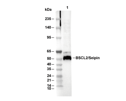 Lane 1: 293T