
|
受注:045-509-1970 |
技術サポート:tech@selleck.co.jp 平日9:00〜18:00 1営業日以内にご連絡を差し上げます |
生物学的記述
| Specificity | EGR2 Antibody (Rabbit mAb) [E12D8] detects endogenous levels of total EGR2 protein. |
|---|---|
| Background | EGR2, also known as KROX20, is a zinc-finger transcription factor that functions as the pivotal master regulator controlling the transition of Schwann cells from the promyelinating stage into active myelin production during peripheral nervous system development. EGR2 expression is itself induced at this developmental transition through a myelinating Schwann cell enhancer element located downstream of the transcriptional start site, and activation of this enhancer requires synergistic cooperation between the POU-domain transcription factors Oct6 and Brn2 and the high-mobility-group transcription factor Sox10, with each factor required to bind its own DNA site independently rather than through a shared composite element; Sox10 additionally engages multiple, largely monomeric binding sites within this enhancer in a configuration that renders EGR2 induction highly sensitive to Sox10 dosage. Once expressed, EGR2 drives the myelination transcriptional program by binding directly to regulatory elements of major myelin genes, including an intronic element of myelin protein zero, and this binding requires cooperative synergy with Sox10 acting through adjacent binding sites within the same regulatory element, a configuration also found in the connexin 32, myelin basic protein, and myelin-associated glycoprotein genes. EGR2 activity is negatively regulated through direct interaction with corepressors of the NAB family, which recruit the NuRD chromatin remodeling complex to attenuate EGR2-driven transactivation, providing a feedback mechanism that tunes the magnitude of the myelination transcriptional response rather than allowing unrestrained target gene activation. Dominant human EGR2 mutations causing peripheral myelinopathies cluster within all three zinc fingers of the DNA-binding domain and generally impair EGR2's capacity to bind DNA, while a separate recessive mutation disrupts the NAB-binding domain specifically, and functional analysis of neuropathy-associated EGR2 mutants shows that at least a subset does not impair EGR2's own DNA binding but instead selectively attenuates recruitment of Sox10 to the shared regulatory element, uncoupling the cooperative EGR2-Sox10 mechanism required for full myelin protein zero induction. A separate germline mutation affecting isoleucine 268, which specifically disrupts the EGR2-NAB interaction without affecting DNA binding, produces congenital hypomyelinating neuropathy with a biphasic disease course in mouse models, directly demonstrating that loss of NAB-mediated feedback repression, independent of any defect in EGR2 DNA-binding capacity, is sufficient to cause peripheral myelin pathology. |
使用情報
| Application | WB | Dilution |
|
||
|---|---|---|---|---|---|
| Reactivity | Human | ||||
| Source | Rabbit Monoclonal Antibody | MW | 50 kDa | ||
| Storage Buffer | PBS, pH 7.2+50% Glycerol+0.05% BSA+0.01% NaN3 | Storage (from the date of receipt) |
-20°C (avoid freeze-thaw cycles), 2 years | ||
| WB |
Experimental Protocol:
Sample preparation
1. Tissue: Lyse the tissue sample by adding an appropriate volume of ice-cold RIPA/Nuclear Lysis Buffer (containing Protease Inhibitor Cocktail),and homogenize the tissue at a low temperature or lyse it by sonication on ice, then incubate on ice for 30 minutes. 2. Adherent cell: Aspirate the culture medium and wash the cells with ice-cold PBS twice. Lyse the cells by adding an appropriate volume of RIPA/Nuclear Lysis Buffer (containing Protease Inhibitor Cocktail) , sonicate to lyse the cells, and incubate on ice for 30 minutes. 3. Suspension cell: Transfer the culture medium to a pre-cooled centrifuge tube. Centrifuge and aspirate the supernatant. Wash the cells with ice-cold PBS twice. Lyse the cells by adding an appropriate volume of RIPA/Nuclear Lysis Buffer (containing Protease Inhibitor Cocktail) , sonicate to lyse the cells, and incubate on ice for 30 minutes. 4. Place the lysate into a pre-cooled microcentrifuge tube. Centrifuge at 4°C for 15 min. Collect the supernatant;
5. Remove a small volume of lysate to determine the protein concentration;
6. Combine the lysate with protein loading buffer. Boil 20 µL sample under 95-100°C for 5 min. Centrifuge for 5 min after cool down on ice.
Electrophoretic separation
1. According to the concentration of extracted protein, load appropriate amount of protein sample and marker onto SDS-PAGE gels for electrophoresis. Recommended separating gel (lower gel) concentration: 10%. Reference Table for Selecting SDS-PAGE Separation Gel Concentrations 2. Power up 80V for 30 minutes. Then the power supply is adjusted (110 V~150 V), the Marker is observed, and the electrophoresis can be stopped when the indicator band of the predyed protein Marker where the protein is located is properly separated. (Note that the current should not be too large when electrophoresis, too large current (more than 150 mA) will cause the temperature to rise, affecting the result of running glue. If high currents cannot be avoided, an ice bath can be used to cool the bath.)
Transfer membrane
1. Take out the converter, soak the clip and consumables in the pre-cooled converter;
2. Activate PVDF membrane with methanol for 1 min and rinse with transfer buffer;
3. Install it in the order of "black edge of clip - sponge - filter paper - filter paper - glue -PVDF membrane - filter paper - filter paper - sponge - white edge of clip"; 4. The protein was electrotransferred to PVDF membrane. ( 0.45 µm PVDF membrane is recommended ) Reference Table for Selecting PVDF Membrane Pore Size Specifications Recommended conditions for wet transfer: 200 mA, 120 min. ( Note that the transfer conditions can be adjusted according to the protein size. For high-molecular-weight proteins, a higher current and longer transfer time are recommended. However, ensure that the transfer tank remains at a low temperature to prevent gel melting.)
Block
1. After electrotransfer, wash the film with TBST at room temperature for 5 minutes;
2. Incubate the film in the blocking solution for 1 hour at room temperature;
3. Wash the film with TBST for 3 times, 5 minutes each time.
Antibody incubation
1. Use primary antibody dilution buffer to prepare the primary antibody working liquid (recommended dilution ratio for primary antibody 1:1000), gently shake and incubate with the film at 4°C overnight; 2. Wash the film with TBST 3 times, 5 minutes each time;
3. Add the secondary antibody to the blocking solution and incubate with the film gently at room temperature for 1 hour;
4. After incubation, wash the film with TBST 3 times for 5 minutes each time.
Antibody staining
1. Add the prepared ECL luminescent substrate (or select other color developing substrate according to the second antibody) and mix evenly;
2. Incubate with the film for 1 minute, remove excess substrate (keep the film moist), wrap with plastic film, and expose in the imaging system.
|
References
|
Application Data
WB
Validated by Selleck
-
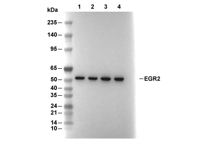 Lane 1: LnCap, Lane 2: HepG2, Lane 3: MCF7, Lane 4: SH-SY5Y