|
受注:045-509-1970 |
技術サポート:tech@selleck.co.jp 平日9:00〜18:00 1営業日以内にご連絡を差し上げます |
生物学的記述
| Specificity | EphB2 Antibody [D9P3] detects endogenous levels of total EphB2 protein. |
|---|---|
| Background | Eph receptor B2 (EphB2) is a transmembrane receptor tyrosine kinase of the EphB subfamily that recognizes membrane‑anchored ephrin‑B ligands, primarily ephrin‑B2 and ephrin‑B3, to mediate contact‑dependent bidirectional signaling that organizes cell positioning, boundary formation, axon guidance, vascular patterning, and tissue remodeling in development and disease. The receptor ectodomain comprises a ligand‑binding domain, a cysteine‑rich region, and two fibronectin type III repeats, followed by a single transmembrane helix and an intracellular segment with a juxtamembrane regulatory region, a tyrosine kinase domain, a sterile‑α‑motif (SAM) domain, and a PDZ‑binding motif; recent crystallography of the full ectodomain shows that EphB2 forms homotypic head‑to‑tail interactions between its ligand‑binding and fibronectin domains that create autoinhibitory receptor–receptor assemblies, with ephrin binding and post‑translational modifications modulating this arrangement to permit higher‑order clustering required for robust downstream signaling. Ligand engagement at sites of cell–cell contact induces EphB2 clustering, activation‑loop phosphorylation, and recruitment of SH2/SH3‑containing effectors including Src‑family kinases, p120RasGAP, Nck, and focal adhesion kinase; through these complexes EphB2 regulates small GTPases of the Rho and Ras families and can either repress or stabilize Ras–ERK MAPK signaling depending on the composition of its phosphotyrosine motifs and SH2 docking sites, such that engineered alterations in RasGAP versus Grb2 binding within EphB2 shift ERK output and correlate with neurite retraction versus extension, linking MAPK regulation directly to growth cone collapse and axon guidance decisions. EphB2 forward signaling also intersects with PI3K–Akt, JNK, and Src–FAK pathways to modulate cytoskeletal dynamics, adhesion complex turnover, and cell migration, while ephrin‑B reverse signaling through its own cytoplasmic tail and PDZ‑protein interactions coordinates complementary responses in the ligand‑expressing cell, creating a bidirectional code that governs cell sorting, boundary sharpening, and morphogenetic movements in the nervous and vascular systems. In the adult brain, EphB2 is enriched at excitatory synapses where it forms complexes with NMDA receptors and scaffolding proteins, potentiates synaptic NMDAR function, and regulates spine morphogenesis and synaptic plasticity, and restoration of EphB2 expression in an Alzheimer’s disease mouse model rescues NMDAR‑dependent long‑term potentiation and spatial memory, implicating loss of EphB2‑NMDAR signaling as a contributing mechanism in cognitive decline. In cancer, EphB2 expression is frequently dysregulated and exerts context‑dependent tumor‑promoting or tumor‑suppressive functions: elevated EphB2 is reported in colorectal, gastric, hepatic, and breast carcinomas, where it can enhance proliferation, survival, autophagy, invasion, and epithelial–mesenchymal transition through sustained activation of oncogenic pathways and remodeling of the tumor microenvironment, yet in certain settings compartmentalized EphB2 signaling at tumor–stroma boundaries restricts cell intermingling and may suppress early tumor expansion, reflecting the dualistic nature of Eph receptor signaling. EphB2 is also upregulated in fibrotic liver and kidney, where it promotes activation of hepatic stellate cells and myofibroblast differentiation and contributes to deposition of extracellular matrix, positioning EphB2 as a driver of organ fibrosis and a candidate therapeutic target in chronic liver and kidney disease. |
使用情報
| Application | WB, IHC | Dilution |
|
||||
|---|---|---|---|---|---|---|---|
| Reactivity | Mouse, Rat, Human | ||||||
| Source | Rabbit Monoclonal Antibody | MW | 117 kDa | ||||
| Storage Buffer | PBS, pH 7.2+50% Glycerol+0.05% BSA+0.01% NaN3 | Storage (from the date of receipt) |
-20°C (avoid freeze-thaw cycles), 2 years | ||||
References
|
Application Data
WB
Validated by Selleck
-
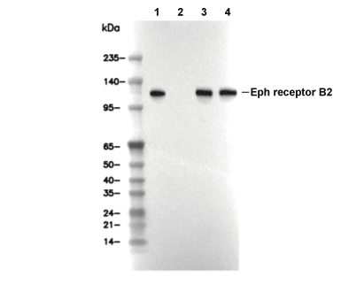 Lane 1: HCT116, Lane 2: HCT116 (KO EPHB2), Lane 3: Mouse brain, Lane 4: Rat brain