
|
受注:045-509-1970 |
技術サポート:tech@selleck.co.jp 平日9:00〜18:00 1営業日以内にご連絡を差し上げます |
生物学的記述
| Specificity | FKBP5 Antibody (Rabbit mAb) [J11H16] detects endogenous levels of total FKBP5 protein. |
|---|---|
| Background | FKBP5, also termed FKBP51, is an immunophilin family member with peptidyl‑prolyl isomerase activity and co‑chaperone functions that modulate steroid hormone receptor signaling, stress‑axis regulation, and kinase pathways at the interface of endocrine and inflammatory control. The protein contains an N‑terminal FK1 domain with cis–trans prolyl isomerase and FK506/rapamycin‑binding activity, an FK2‑like domain with mainly scaffolding function, and a C‑terminal region bearing tetratricopeptide repeat motifs that mediate binding to HSP90 and assembly into multichaperone complexes with glucocorticoid, progesterone, androgen, and mineralocorticoid receptors. FKBP5 associates with HSP90‑bound glucocorticoid receptor (GR) complexes and reduces ligand affinity and efficiency of GR nuclear translocation, resulting in a receptor state with lower hormone sensitivity; GR activation then induces FKBP5 transcription via intronic glucocorticoid response elements, creating an ultra‑short negative feedback loop on GR signaling and hypothalamic–pituitary–adrenal (HPA) axis feedback. This autoregulatory circuit influences systemic cortisol dynamics, as alleles associated with stronger FKBP5 upregulation after GR activation correlate with increased GR resistance, reduced feedback inhibition, and prolonged stress‑hormone elevation in humans. FKBP5 also functions as a scaffold in kinase signaling: it binds AKT through its FK1 domain and the phosphatase PHLPP via TPR motifs, promotes their interaction, and facilitates dephosphorylation of AKT at Ser473, thereby constraining AKT activity and linking FKBP5 abundance to PI3K–Akt pathway output and cell survival signaling. At the level of inflammatory and cardiovascular control, FKBP5 expression is epigenetically and transcriptionally upregulated by aging and chronic stress, enhances NF‑κB–related signaling, and associates with a pro‑inflammatory profile and increased cardiovascular risk, indicating that FKBP5 connects stress endocrine mechanisms to innate immune activation and vascular pathology. Genetic and epigenetic variation at the FKBP5 locus, including functional polymorphisms and stress‑responsive DNA methylation changes, associates with altered FKBP5 inducibility, HPA‑axis reactivity, and susceptibility to major depression, post‑traumatic stress disorder, bipolar disorder, and related stress‑induced psychiatric conditions, and these same variants can influence response rates to antidepressant therapy. |
使用情報
| Application | WB, IP | Dilution |
|
||||
|---|---|---|---|---|---|---|---|
| Reactivity | Human, Mouse, Rat, Monkey | ||||||
| Source | Rabbit Monoclonal Antibody | MW | 51 kDa | ||||
| Storage Buffer | PBS, pH 7.2+50% Glycerol+0.05% BSA+0.01% NaN3 | Storage (from the date of receipt) |
-20°C (avoid freeze-thaw cycles), 2 years | ||||
| WB |
Experimental Protocol:
Sample preparation
1. Tissue: Lyse the tissue sample by adding an appropriate volume of ice-cold RIPA/NP-40 Lysis Buffer (containing Protease Inhibitor Cocktail),and homogenize the tissue at a low temperature or lyse it by sonication on ice, then incubate on ice for 30 minutes. 2. Adherent cell: Aspirate the culture medium and wash the cells with ice-cold PBS twice. Lyse the cells by adding an appropriate volume of RIPA/NP-40 Lysis Buffer (containing Protease Inhibitor Cocktail) , sonicate to lyse the cells, and incubate on ice for 30 minutes. 3. Suspension cell: Transfer the culture medium to a pre-cooled centrifuge tube. Centrifuge and aspirate the supernatant. Wash the cells with ice-cold PBS twice. Lyse the cells by adding an appropriate volume of RIPA/NP-40 Lysis Buffer (containing Protease Inhibitor Cocktail) , sonicate to lyse the cells, and incubate on ice for 30 minutes. 4. Place the lysate into a pre-cooled microcentrifuge tube. Centrifuge at 4°C for 15 min. Collect the supernatant;
5. Remove a small volume of lysate to determine the protein concentration;
6. Combine the lysate with protein loading buffer. Boil 20 µL sample under 95-100°C for 5 min. Centrifuge for 5 min after cool down on ice.
Electrophoretic separation
1. According to the concentration of extracted protein, load appropriate amount of protein sample and marker onto SDS-PAGE gels for electrophoresis. Recommended separating gel (lower gel) concentration: 10%. Reference Table for Selecting SDS-PAGE Separation Gel Concentrations 2. Power up 80V for 30 minutes. Then the power supply is adjusted (110 V~150 V), the Marker is observed, and the electrophoresis can be stopped when the indicator band of the predyed protein Marker where the protein is located is properly separated. (Note that the current should not be too large when electrophoresis, too large current (more than 150 mA) will cause the temperature to rise, affecting the result of running glue. If high currents cannot be avoided, an ice bath can be used to cool the bath.)
Transfer membrane
1. Take out the converter, soak the clip and consumables in the pre-cooled converter;
2. Activate PVDF membrane with methanol for 1 min and rinse with transfer buffer;
3. Install it in the order of "black edge of clip - sponge - filter paper - filter paper - glue -PVDF membrane - filter paper - filter paper - sponge - white edge of clip"; 4. The protein was electrotransferred to PVDF membrane. ( 0.45 µm PVDF membrane is recommended ) Reference Table for Selecting PVDF Membrane Pore Size Specifications Recommended conditions for wet transfer: 200 mA, 120 min. ( Note that the transfer conditions can be adjusted according to the protein size. For high-molecular-weight proteins, a higher current and longer transfer time are recommended. However, ensure that the transfer tank remains at a low temperature to prevent gel melting.)
Block
1. After electrotransfer, wash the film with TBST at room temperature for 5 minutes;
2. Incubate the film in the blocking solution for 1 hour at room temperature;
3. Wash the film with TBST for 3 times, 5 minutes each time.
Antibody incubation
1. Use primary antibody dilution buffer to prepare the primary antibody working liquid (recommended dilution ratio for primary antibody 1:1000), gently shake and incubate with the film at 4°C overnight; 2. Wash the film with TBST 3 times, 5 minutes each time;
3. Add the secondary antibody to the blocking solution and incubate with the film gently at room temperature for 1 hour;
4. After incubation, wash the film with TBST 3 times for 5 minutes each time.
Antibody staining
1. Add the prepared ECL luminescent substrate (or select other color developing substrate according to the second antibody) and mix evenly;
2. Incubate with the film for 1 minute, remove excess substrate (keep the film moist), wrap with plastic film, and expose in the imaging system.
|
References
|
Application Data
WB
Validated by Selleck
-
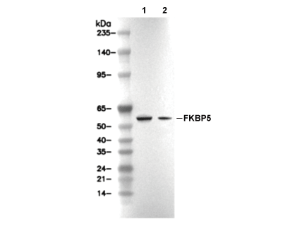 Lane 1: HT1080, Lane 2: Hela