|
受注:045-509-1970 |
技術サポート:tech@selleck.co.jp 平日9:00〜18:00 1営業日以内にご連絡を差し上げます |
生物学的記述
| Specificity | GPNMB Antibody [J15J20] detects endogenous levels of total GPNMB protein. |
|---|---|
| Background | GPNMB, or glycoprotein non-metastatic melanoma protein B, belongs to the type I transmembrane glycoprotein family and serves as a multifaceted regulator of cellular adhesion, migration, and lysosomal dynamics across diverse tissues. It features an N-terminal polycystic kidney disease-like domain, an RGD integrin-binding motif, a cleaved ectodomain, and a short cytoplasmic tail with a hemITAM motif that recruits signaling adaptors. GPNMB localizes primarily to intracellular compartments in normal cells but translocates to the plasma membrane in cancer, where ADAM10-mediated ectodomain shedding releases soluble GPNMB that binds endothelial integrins to trigger VEGF/NRP-1 signaling, FAK activation, and directed migration essential for angiogenesis; full-length GPNMB engages α5β1 integrin via its RGD motif to drive PI3K/AKT/mTOR and ERK/MAPK cascades that upregulate MMP-2/9 for matrix remodeling and ZEB1 for epithelial-mesenchymal transition. GPNMB silencing suppresses proliferation, invasion, and tube formation by downregulating MMP-3 and HIF1α while elevating apoptosis, revealing its central role in tumor-stroma crosstalk where shed ectodomain recruits endothelial support for vascular mimicry in glioma and breast cancer cells. GPNMB governs melanosome transfer to keratinocytes, osteoblast/osteoclast differentiation through RANKL modulation, and dendritic cell maturation via adhesion to VCAM-1, positioning it as a prime target for researchers dissecting lysosomal biogenesis or immune cell trafficking in tissue repair models. Its expression in skin, heart, kidney, and muscle supports homeostasis through autophagy-lysosome flux and debris clearance, with tissue-specific shedding controlled by HB-EGF/EGFR phosphorylation. Dysregulation through overexpression correlates with metastasis in melanoma, glioma, and triple-negative breast cancer, where surface localization predicts poor outcome. |
使用情報
| Application | WB, IP, IHC | Dilution |
|
||||||
|---|---|---|---|---|---|---|---|---|---|
| Reactivity | Human | ||||||||
| Source | Rabbit Monoclonal Antibody | MW | 95 kDa, 120 kDa | ||||||
| Storage Buffer | PBS, pH 7.2+50% Glycerol+0.05% BSA+0.01% NaN3 | Storage (from the date of receipt) |
-20°C (avoid freeze-thaw cycles), 2 years | ||||||
References
|
Application Data
WB
Validated by Selleck
-
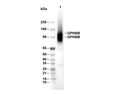 Lane 1: SK-BR-3