|
受注:045-509-1970 |
技術サポート:tech@selleck.co.jp 平日9:00〜18:00 1営業日以内にご連絡を差し上げます |
生物学的記述
| Specificity | Granzyme B Antibody [A1B9] detects endogenous levels of total Granzyme B protein. |
|---|---|
| Background | Granzyme B is a tryptase‑type serine protease of the granzyme family that is predominantly expressed by cytotoxic T lymphocytes and natural killer cells, where it serves as a major effector of perforin‑dependent cell killing in response to infected or transformed targets. It is synthesized as a glycosylated zymogen that is processed into its mature active form within cytotoxic granules, and its activity is maintained at neutral pH, which prevents premature apoptosis inside the killer cell. Upon recognition of a target, cytotoxic lymphocytes polarize their granules toward the immunological synapse and release perforin‑containing vesicles; perforin forms pores in the target‑cell membrane that allow granzyme B to enter the cytosol and, in some contexts, the nucleus, where it cleaves a broad spectrum of aspartate‑containing substrates. Granzyme B directly activates procaspases, especially caspase‑3 and its downstream targets, and can also engage the mitochondrial pathway by cleaving the pro‑apoptotic Bcl‑2 family protein Bid, which in turn promotes Bax/Bak‑mediated cytochrome‑c release and apoptosome assembly, thereby amplifying caspase‑dependent cell death. Granzyme B cleaves nuclear and cytosolic factors involved in DNA integrity and repair, such as ICAD and PARP, and can induce caspase‑independent apoptotic‑like events by targeting structural and regulatory proteins. Granzyme B‑dependent killing is essential for antiviral CD8+ T‑cell responses and tumor surveillance, and dysregulated granzyme B expression or extracellular release has been linked to autoinflammatory and autoimmune conditions. Granzyme B integrates signals from the perforin pathway, caspase activation, and mitochondrial dysfunction, and it is frequently used as a mechanistic marker and potential therapeutic target in studies of immune‑mediated cell death and immunotherapy. |
使用情報
| Application | WB, IHC, FCM | Dilution |
|
||||||
|---|---|---|---|---|---|---|---|---|---|
| Reactivity | Mouse | ||||||||
| Source | Rabbit Monoclonal Antibody | MW | 30 kDa | ||||||
| Storage Buffer | PBS, pH 7.2+50% Glycerol+0.05% BSA+0.01% NaN3 | Storage (from the date of receipt) |
-20°C (avoid freeze-thaw cycles), 2 years | ||||||
References
|
Application Data
WB
Validated by Selleck
-
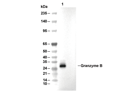 Lane 1: Mouse CD8+T Cells