|
受注:045-509-1970 |
技術サポート:tech@selleck.co.jp 平日9:00〜18:00 1営業日以内にご連絡を差し上げます |
生物学的記述
| Specificity | Histone H3 (mono methyl Lys27) Antibody [G24J14] recognizes endogenous levels of histone H3 protein only when mono-methylated at Lys27. |
|---|---|
| Background | Histone H3 monomethyl Lys27 (H3K27me1) is a pivotal post-translational modification that adds a methyl group to the ε-amino group of Lys27 on histone H3, a core nucleosomal protein forming octamers with H2A, H2B, and H4 to package DNA. This modification occurs on the flexible N-terminal tail (amino acids 1–40) and the globular histone fold (amino acids 94–123), and is deposited by PRC2/EZH2 (which prefers mono- over tri-methylation) or through SETD2-opposed pathways. H3K27me1 marks permissive, active, or poised chromatin at gene bodies, enhancers, and bivalent promoters during development and differentiation, in contrast to the repressive H3K27me3 mark. The methylated K27 embeds in a hydrophobic H3-H3 interface pocket (modulated by Ala28 and Pro30), can coexist with H3K36me3 to enhance nucleosome mobility and RNA Pol II access (unlike the compact PRC1-bound H3K27me3 state), recruits reader proteins via PHD or Tudor motifs (such as BPTF, but not CBX chromodomains), and is reversible by JMJD3/UTX demethylases (primarily active on di- and tri-methylation, but also on mono-methylation). H3K27me1 drives transcriptional activation and elongation, enhancer-promoter looping, high gene expression, genome programming during embryogenesis and erythroid differentiation, and antagonism of H3K27me3 at lineage-specific loci. Dysregulation of H3K27me1, such as through PRC2 mutations that shift the mono- to tri-methylation balance, underlies cancers (including leukemia and pediatric tumors) and developmental disorders; treatment with EZH2 inhibitors can elevate H3K27me1 and activate tumor suppressor genes. |
使用情報
| Application | WB, IP | Dilution |
|
||||
|---|---|---|---|---|---|---|---|
| Reactivity | Human, Mouse, Rat, Monkey | ||||||
| Source | Rabbit Monoclonal Antibody | MW | 17 kDa | ||||
| Storage Buffer | PBS, pH 7.2+50% Glycerol+0.05% BSA+0.01% NaN3 | Storage (from the date of receipt) |
-20°C (avoid freeze-thaw cycles), 2 years | ||||
| WB |
Experimental Protocol:
Sample preparation
1. Tissue: Lyse the tissue sample by adding an appropriate volume of ice-cold RIPA/Nuclear Lysis Buffer (containing Protease Inhibitor Cocktail),and homogenize the tissue at a low temperature. 2. Adherent cell: Aspirate the culture medium and wash the cells with ice-cold PBS twice. Lyse the cells by adding an appropriate volume of RIPA/Nuclear Lysis Buffer (containing Protease Inhibitor Cocktail) and put the sample on ice for 5 min. 3. Suspension cell: Transfer the culture medium to a pre-cooled centrifuge tube. Centrifuge and aspirate the supernatant. Wash the cells with ice-cold PBS twice. Lyse the cells by adding an appropriate volume of RIPA/Nuclear Lysis Buffer (containing Protease Inhibitor Cocktail) and put the sample on ice for 5 min. 4. Place the lysate into a pre-cooled microcentrifuge tube. Centrifuge at 4°C for 15 min. Collect the supernatant;
5. Remove a small volume of lysate to determine the protein concentration;
6. Combine the lysate with protein loading buffer. Boil 20 µL sample under 95-100°C for 5 min. Centrifuge for 5 min after cool down on ice.
Electrophoretic separation
1. According to the concentration of extracted protein, load appropriate amount of protein sample and marker onto SDS-PAGE gels for electrophoresis. Recommended separating gel (lower gel) concentration: 10%. Reference Table for Selecting SDS-PAGE Separation Gel Concentrations 2. Power up 80V for 30 minutes. Then the power supply is adjusted (110 V~150 V), the Marker is observed, and the electrophoresis can be stopped when the indicator band of the predyed protein Marker where the protein is located is properly separated. (Note that the current should not be too large when electrophoresis, too large current (more than 150 mA) will cause the temperature to rise, affecting the result of running glue. If high currents cannot be avoided, an ice bath can be used to cool the bath.)
Transfer membrane
1. Take out the converter, soak the clip and consumables in the pre-cooled converter;
2. Activate PVDF membrane with methanol for 1 min and rinse with transfer buffer;
3. Install it in the order of "black edge of clip - sponge - filter paper - filter paper - glue -PVDF membrane - filter paper - filter paper - sponge - white edge of clip"; 4. The protein was electrotransferred to PVDF membrane. ( 0.22 µm PVDF membrane is recommended )) Reference Table for Selecting PVDF Membrane Pore Size Specifications Recommended conditions for wet transfer: 200 mA, 60 min. ( Note that the transfer conditions can be adjusted according to the protein size. For high-molecular-weight proteins, a higher current and longer transfer time are recommended. However, ensure that the transfer tank remains at a low temperature to prevent gel melting.)
Block
1. After electrotransfer, wash the film with TBST at room temperature for 5 minutes;
2. Incubate the film in the blocking solution for 1 hour at room temperature;
3. Wash the film with TBST for 3 times, 5 minutes each time.
Antibody incubation
1. Use 5% skim milk powder to prepare the primary antibody working liquid (recommended dilution ratio for primary antibody 1:1000), gently shake and incubate with the film at 4°C overnight; 2. Wash the film with TBST 3 times, 5 minutes each time;
3. Add the secondary antibody to the blocking solution and incubate with the film gently at room temperature for 1 hour;
4. After incubation, wash the film with TBST 3 times for 5 minutes each time.
Antibody staining
1. Add the prepared ECL luminescent substrate (or select other color developing substrate according to the second antibody) and mix evenly;
2. Incubate with the film for 1 minute, remove excess substrate (keep the film moist), wrap with plastic film, and expose in the imaging system.
|
References
|
Application Data
WB
Validated by Selleck
-
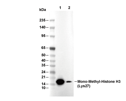 Lane 1: Hela, Lane 2: C2C12