
|
受注:045-509-1970 |
技術サポート:tech@selleck.co.jp 平日9:00〜18:00 1営業日以内にご連絡を差し上げます |
生物学的記述
| Specificity | HOXA9 Antibody (Rabbit mAb) [A6N14] detects endogenous levels of total HOXA9 protein. |
|---|---|
| Background | HOXA9 is a homeobox family transcription factor that contains a conserved homeodomain for sequence‑specific DNA binding and is expressed at high levels in hematopoietic stem and progenitor cells, where it regulates gene expression programs that support stem cell expansion, lineage commitment and early blood cell development. The protein functions within HOX clusters and forms cooperative complexes with TALE cofactors such as PBX and MEIS, using its homeodomain and flanking regions to bind regulatory elements and control a broad transcriptional network that includes genes involved in self‑renewal, survival, adhesion and cytokine signaling, and its expression decreases as progenitors differentiate into mature hematopoietic lineages. In human embryonic stem cell differentiation, HOXA9 expression parallels hematopoietic progression and is restricted to hemogenic precursors; gain‑ and loss‑of‑function studies show that HOXA9 enhances commitment of these precursors into primitive and total CD45‑positive blood cells, partly in collaboration with NF‑κB signaling, indicating that HOXA9 integrates developmental cues with transcriptional control of early hematopoietic specification. In leukemia, HOXA9 is frequently deregulated, particularly in acute myeloid leukemia with MLL rearrangements and other upstream lesions, and sustained HOXA9 expression maintains gene expression for multiple anti‑apoptotic pathways and growth programs; HOXA9 overexpression in myeloid progenitors enforces a stem‑like transcriptional state, while combined expression with MEIS1 or PBX1 drives full leukemic transformation, establishing HOXA9 as a central effector of leukemogenic gene expression. Mechanistically, HOXA9 reprograms the enhancer landscape in myeloid and B‑cell progenitors by binding noncoding regulatory sequences and recruiting C/EBPα and the MLL3/MLL4 methyltransferase complex, which leads to formation of leukemia‑specific enhancers, activation of an ectopic embryonic gene program and maintenance of stemness and survival genes that support malignant progression. Transcriptome analyses in human hematopoietic cells show that enforced HOXA9 expression upregulates clusters of genes associated with stem cell maintenance, cell‑cycle progression and resistance to apoptosis, while HOXA9 deficiency impairs normal hematopoietic development and reduces repopulating capacity, confirming its dual role as a positive regulator of physiological stem cell function and, when dysregulated, a driver of leukemia. In solid tumors, HOXA9 is aberrantly expressed in multiple cancer types and functions as a transcriptional regulator of EMT, autophagy, cell‑cycle, metabolic and major signaling pathways, including Wnt, TGF‑β, PI3K/AKT, AMPK and NF‑κB, through PBX/MEIS‑mediated binding to promoters and enhancers of these pathway components, and pan‑cancer analyses indicate that HOXA9 expression patterns have diagnostic and prognostic significance linked to tumor stage and immune infiltration. Across hematologic and solid malignancies, HOXA9 thus behaves as a modular transcription factor whose homeodomain‑based DNA binding and cofactor interactions remodel enhancer and promoter landscapes, sustain anti‑apoptotic and stemness‑related gene expression, and couple developmental signaling pathways to oncogenic programs. |
使用情報
| Application | WB, IP | Dilution |
|
||||
|---|---|---|---|---|---|---|---|
| Reactivity | Human | ||||||
| Source | Rabbit Monoclonal Antibody | MW | 30 kDa | ||||
| Storage Buffer | PBS, pH 7.2+50% Glycerol+0.05% BSA+0.01% NaN3 | Storage (from the date of receipt) |
-20°C (avoid freeze-thaw cycles), 2 years | ||||
| WB |
Experimental Protocol:
Sample preparation
1. Tissue: Lyse the tissue sample by adding an appropriate volume of ice-cold RIPA/NP-40 Lysis Buffer (containing Protease Inhibitor Cocktail),and homogenize the tissue at a low temperature or lyse it by sonication on ice, then incubate on ice for 30 minutes. 2. Adherent cell: Aspirate the culture medium and wash the cells with ice-cold PBS twice. Lyse the cells by adding an appropriate volume of RIPA/NP-40 Lysis Buffer (containing Protease Inhibitor Cocktail) , sonicate to lyse the cells, and incubate on ice for 30 minutes. 3. Suspension cell: Transfer the culture medium to a pre-cooled centrifuge tube. Centrifuge and aspirate the supernatant. Wash the cells with ice-cold PBS twice. Lyse the cells by adding an appropriate volume of RIPA/NP-40 Lysis Buffer (containing Protease Inhibitor Cocktail) , sonicate to lyse the cells, and incubate on ice for 30 minutes. 4. Place the lysate into a pre-cooled microcentrifuge tube. Centrifuge at 4°C for 15 min. Collect the supernatant;
5. Remove a small volume of lysate to determine the protein concentration;
6. Combine the lysate with protein loading buffer. Boil 20 µL sample under 95-100°C for 5 min. Centrifuge for 5 min after cool down on ice.
Electrophoretic separation
1. According to the concentration of extracted protein, load appropriate amount of protein sample and marker onto SDS-PAGE gels for electrophoresis. Recommended separating gel (lower gel) concentration: 10%. Reference Table for Selecting SDS-PAGE Separation Gel Concentrations 2. Power up 80V for 30 minutes. Then the power supply is adjusted (110 V~150 V), the Marker is observed, and the electrophoresis can be stopped when the indicator band of the predyed protein Marker where the protein is located is properly separated. (Note that the current should not be too large when electrophoresis, too large current (more than 150 mA) will cause the temperature to rise, affecting the result of running glue. If high currents cannot be avoided, an ice bath can be used to cool the bath.)
Transfer membrane
1. Take out the converter, soak the clip and consumables in the pre-cooled converter;
2. Activate PVDF membrane with methanol for 1 min and rinse with transfer buffer;
3. Install it in the order of "black edge of clip - sponge - filter paper - filter paper - glue -PVDF membrane - filter paper - filter paper - sponge - white edge of clip"; 4. The protein was electrotransferred to PVDF membrane. ( 0.45 µm PVDF membrane is recommended ) Reference Table for Selecting PVDF Membrane Pore Size Specifications Recommended conditions for wet transfer: 200 mA, 60 min. ( Note that the transfer conditions can be adjusted according to the protein size. For high-molecular-weight proteins, a higher current and longer transfer time are recommended. However, ensure that the transfer tank remains at a low temperature to prevent gel melting.)
Block
1. After electrotransfer, wash the film with TBST at room temperature for 5 minutes;
2. Incubate the film in the blocking solution for 1 hour at room temperature;
3. Wash the film with TBST for 3 times, 5 minutes each time.
Antibody incubation
1. Use primary antibody dilution buffer to prepare the primary antibody working liquid (recommended dilution ratio for primary antibody 1:1000), gently shake and incubate with the film at 4°C overnight; 2. Wash the film with TBST 3 times, 5 minutes each time;
3. Add the secondary antibody to the blocking solution and incubate with the film gently at room temperature for 1 hour;
4. After incubation, wash the film with TBST 3 times for 5 minutes each time.
Antibody staining
1. Add the prepared ECL luminescent substrate (or select other color developing substrate according to the second antibody) and mix evenly;
2. Incubate with the film for 1 minute, remove excess substrate (keep the film moist), wrap with plastic film, and expose in the imaging system.
|
References
|
Application Data
WB
Validated by Selleck
-
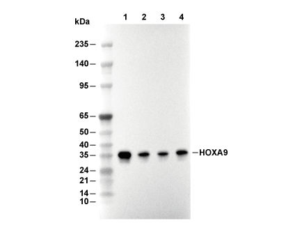 Lane 1: HepG2, Lane 2: U937, Lane 3: SW480, Lane 4: 293T