
|
受注:045-509-1970 |
技術サポート:tech@selleck.co.jp 平日9:00〜18:00 1営業日以内にご連絡を差し上げます |
生物学的記述
| Specificity | IRF-7 Antibody (Rabbit mAb) [C15N21] detects endogenous levels of total IRF-7 protein. |
|---|---|
| Background | Interferon regulatory factor 7 (IRF-7) is a member of the IRF transcription factor family that binds specific DNA elements in interferon-stimulated promoters and integrates upstream innate immune receptor signaling into type I and III interferon gene expression, especially in plasmacytoid dendritic cells and lymphoid lineages, where its expression is enriched and inducible by virus, lipopolysaccharide, and type I interferons. The N-terminal region contains a conserved helix–turn–helix DNA-binding domain that recognizes interferon regulatory elements, while the C-terminal regulatory region carries multiple serine-rich motifs and interaction surfaces that control dimerization, nuclear import, and cofactor recruitment. Upstream activation begins at pattern-recognition receptors such as endosomal Toll-like receptors and cytosolic nucleic acid sensors, which signal through adaptor proteins including MyD88 or TRIF, TRAF family ubiquitin ligases, and TANK-binding kinase 1 or IKK-related kinases that phosphorylate IRF-7 on clustered serine residues in the C terminus. Phosphorylated IRF-7 forms homodimers or heterodimers with other IRFs, undergoes conformational exposure of its nuclear localization signal, accumulates in the nucleus, and assembles on interferon promoters together with chromatin-modifying coactivators to drive a broad antiviral transcriptional program. This transcriptional output reinforces interferon signaling through a positive feedback loop, shapes the amplitude and duration of type I interferon waves, and coordinates expression of numerous interferon-stimulated genes that regulate viral replication, antigen presentation, and inflammatory mediator production. Multiple post-translational modifications fine-tune this activity: phosphorylation marks correlate with activation, K63-linked ubiquitination promotes full transactivation, and other ubiquitin linkages or proteasomal targeting limit IRF-7 abundance and prevent excessive interferon output. IRF-7 also interfaces with NF-κB and AP-1 pathways at shared target promoters and participates in Epstein–Barr virus latency programs, where its induction and activation by latent membrane protein 1 link viral oncogenic signaling to interferon-related transcriptional networks. In physiological immunity, IRF-7 is critical for early systemic antiviral defense, shaping plasmacytoid dendritic cell function, influencing B cell responses, and contributing to broader orchestration of innate and adaptive immune communication. Dysregulated IRF-7 expression or activity alters interferon production thresholds, associates with autoimmune phenotypes characterized by chronic type I interferon signatures, and influences susceptibility or progression in infection, cancer, and other inflammation-associated pathologies where interferon balance is a major determinant of tissue outcome. |
使用情報
| Application | WB, IP | Dilution |
|
||||
|---|---|---|---|---|---|---|---|
| Reactivity | Mouse, Rat, Hamster | ||||||
| Source | Rabbit Monoclonal Antibody | MW | 54 kDa | ||||
| Storage Buffer | PBS, pH 7.2+50% Glycerol+0.05% BSA+0.01% NaN3 | Storage (from the date of receipt) |
-20°C (avoid freeze-thaw cycles), 2 years | ||||
| WB |
Experimental Protocol:
Sample preparation
1. Tissue: Lyse the tissue sample by adding an appropriate volume of ice-cold RIPA/NP-40 Lysis Buffer (containing Protease Inhibitor Cocktail),and homogenize the tissue at a low temperature or lyse it by sonication on ice, then incubate on ice for 30 minutes. 2. Adherent cell: Aspirate the culture medium and wash the cells with ice-cold PBS twice. Lyse the cells by adding an appropriate volume of RIPA/NP-40 Lysis Buffer (containing Protease Inhibitor Cocktail) , sonicate to lyse the cells, and incubate on ice for 30 minutes. 3. Suspension cell: Transfer the culture medium to a pre-cooled centrifuge tube. Centrifuge and aspirate the supernatant. Wash the cells with ice-cold PBS twice. Lyse the cells by adding an appropriate volume of RIPA/NP-40 Lysis Buffer (containing Protease Inhibitor Cocktail) , sonicate to lyse the cells, and incubate on ice for 30 minutes. 4. Place the lysate into a pre-cooled microcentrifuge tube. Centrifuge at 4°C for 15 min. Collect the supernatant;
5. Remove a small volume of lysate to determine the protein concentration;
6. Combine the lysate with protein loading buffer. Boil 20 µL sample under 95-100°C for 5 min. Centrifuge for 5 min after cool down on ice.
Electrophoretic separation
1. According to the concentration of extracted protein, load appropriate amount of protein sample and marker onto SDS-PAGE gels for electrophoresis. Recommended separating gel (lower gel) concentration: 10%. Reference Table for Selecting SDS-PAGE Separation Gel Concentrations 2. Power up 80V for 30 minutes. Then the power supply is adjusted (110 V~150 V), the Marker is observed, and the electrophoresis can be stopped when the indicator band of the predyed protein Marker where the protein is located is properly separated. (Note that the current should not be too large when electrophoresis, too large current (more than 150 mA) will cause the temperature to rise, affecting the result of running glue. If high currents cannot be avoided, an ice bath can be used to cool the bath.)
Transfer membrane
1. Take out the converter, soak the clip and consumables in the pre-cooled converter;
2. Activate PVDF membrane with methanol for 1 min and rinse with transfer buffer;
3. Install it in the order of "black edge of clip - sponge - filter paper - filter paper - glue -PVDF membrane - filter paper - filter paper - sponge - white edge of clip"; 4. The protein was electrotransferred to PVDF membrane. ( 0.45 µm PVDF membrane is recommended ) Reference Table for Selecting PVDF Membrane Pore Size Specifications Recommended conditions for wet transfer: 200 mA, 120 min. ( Note that the transfer conditions can be adjusted according to the protein size. For high-molecular-weight proteins, a higher current and longer transfer time are recommended. However, ensure that the transfer tank remains at a low temperature to prevent gel melting.)
Block
1. After electrotransfer, wash the film with TBST at room temperature for 5 minutes;
2. Incubate the film in the blocking solution for 1 hour at room temperature;
3. Wash the film with TBST for 3 times, 5 minutes each time.
Antibody incubation
1. Use 5% skim milk powder to prepare the primary antibody working liquid (recommended dilution ratio for primary antibody 1:1000), gently shake and incubate with the film at 4°C overnight; 2. Wash the film with TBST 3 times, 5 minutes each time;
3. Add the secondary antibody to the blocking solution and incubate with the film gently at room temperature for 1 hour;
4. After incubation, wash the film with TBST 3 times for 5 minutes each time.
Antibody staining
1. Add the prepared ECL luminescent substrate (or select other color developing substrate according to the second antibody) and mix evenly;
2. Incubate with the film for 1 minute, remove excess substrate (keep the film moist), wrap with plastic film, and expose in the imaging system.
|
References
|
Application Data
WB
Validated by Selleck
-
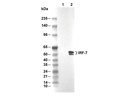 Lane 1: A20, Lane 2: A20 (mIFN-α, 10 ng/ml, 24 h)