|
受注:045-509-1970 |
技術サポート:tech@selleck.co.jp 平日9:00〜18:00 1営業日以内にご連絡を差し上げます |
生物学的記述
| Specificity | LAG3 Antibody [J16E22] detects endogenous levels of total LAG3 protein. |
|---|---|
| Background | Lymphocyte activation gene 3 (LAG‑3, CD223) is a type I transmembrane immune checkpoint receptor of the immunoglobulin superfamily that is expressed on activated CD4 and CD8 T cells, regulatory T cells, natural killer cells, and plasmacytoid dendritic cells, where it serves as a negative regulator of antigen‑driven activation and effector function. The extracellular region is composed of four immunoglobulin‑like domains with overall structural homology to CD4 but contains a distinctive proline‑rich loop within the D1 domain that confers high‑affinity binding to major histocompatibility complex class II molecules, and this extended loop forms the principal contact site for MHC class II on antigen‑presenting cells. The cytoplasmic tail lacks classical inhibitory motifs but carries a unique KIEELE sequence, a conserved serine‑phosphorylation region, and an EP repeat segment that together define non‑canonical signaling modules required for the inhibitory function of LAG‑3 on T cell receptor signaling and cytokine production. Engagement of LAG‑3 with MHC class II dampens proximal TCR signaling and reduces calcium flux, activation of downstream transcriptional programs, and expression of effector cytokines, thereby limiting T cell proliferation and cytotoxicity during sustained antigen exposure. Coexpression with PD‑1 and other co‑inhibitory receptors is frequent on chronically stimulated or exhausted T cells, and functional cooperation between LAG‑3 and PD‑1 accentuates suppression of effector function in the tumor microenvironment and during chronic infection, creating a multilayered checkpoint system that constrains persistent immune activation. Additional ligands such as galectin‑3, LSECtin, and fibrinogen‑like protein 1 engage distinct domains of LAG‑3 and expand its regulatory reach beyond classical MHC class II interactions, connecting this receptor to glycan‑dependent regulation, liver‑resident immune modulation, and tumor‑derived inhibitory signals. Expression of LAG‑3 is induced upon TCR stimulation and maintained by inflammatory cues, while regulatory T cells display constitutive expression that contributes to their suppressive activity, including inhibitory contact with dendritic cells via MHC class II that reduces dendritic‑cell maturation and co‑stimulatory capacity. Elevated or sustained LAG‑3 expression characterizes tumor‑infiltrating lymphocytes across multiple malignancies and is associated with reduced effector function and features of T cell exhaustion, and combined blockade of LAG‑3 with PD‑1 enhances intratumoral T cell activity and underpins current combination immunotherapy strategies. |
使用情報
| Application | WB, IP, IF | Dilution |
|
||||||
|---|---|---|---|---|---|---|---|---|---|
| Reactivity | Mouse | ||||||||
| Source | Rabbit Monoclonal Antibody | MW | 57 kDa | ||||||
| Storage Buffer | PBS, pH 7.2+50% Glycerol+0.05% BSA+0.01% NaN3 | Storage (from the date of receipt) |
-20°C (avoid freeze-thaw cycles), 2 years | ||||||
References
|
Application Data
WB
Validated by Selleck
-
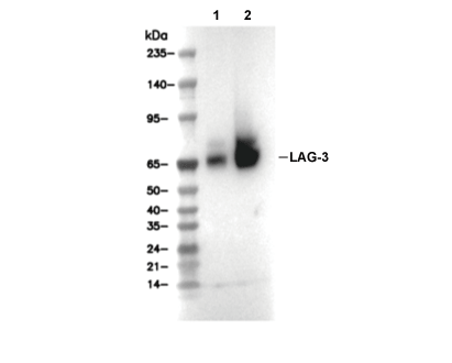 Lane 1: Mouse splenocytes, Lane 2: Mouse splenocytes (Concanavalin A, 5ug/ml, 72 h)