|
受注:045-509-1970 |
技術サポート:tech@selleck.co.jp 平日9:00〜18:00 1営業日以内にご連絡を差し上げます |
生物学的記述
| Specificity | Myosin light chain kinase/MLCK Antibody [F16B16] detects endogenous levels of total Myosin light chain kinase/MLCK protein. |
|---|---|
| Background | Myosin light chain kinase (MLCK, MYLK) is a Ca²⁺/calmodulin-dependent serine/threonine kinase of the myosin kinase family that phosphorylates the regulatory light chain of myosin II and couples Ca²⁺ signals to actomyosin contractility in smooth muscle, skeletal muscle, and nonmuscle cells. The enzyme contains an N‑terminal targeting region that associates with actin–myosin filaments, a central catalytic kinase domain with the consensus HRD motif, and a C‑terminal regulatory segment harboring the calmodulin-binding site and multiple regulatory phosphorylation sites, and this arrangement allows Ca²⁺/calmodulin binding and upstream kinase inputs to control catalytic activity and subcellular positioning. Activation begins when Ca²⁺ binds calmodulin, and the Ca²⁺/calmodulin complex engages the MLCK regulatory domain, relieving autoinhibition and enabling MLCK to phosphorylate the regulatory myosin light chain at a conserved serine, which increases myosin ATPase activity, promotes actin binding, and initiates cross-bridge cycling and force generation in smooth muscle or potentiates force and shortening in skeletal muscle. Smooth muscle–specific MLCK isoforms are further tuned by phosphorylation of residues within the calmodulin-binding region by kinases such as CaMKII, PKA, PKG, PAK, and PKC, which reduce calmodulin affinity and thereby decrease sensitivity to Ca²⁺, while mitogen-activated protein kinases phosphorylate other sites to increase maximal catalytic velocity without changing Ca²⁺ dependence, integrating diverse signaling pathways onto the same contractile effector. MLCK operates in dynamic balance with myosin light chain phosphatase, and the relative activities of these enzymes define the phosphorylation state of myosin light chain, the level of actomyosin interaction, and the presence of latch-like contractile states in smooth muscle. Nonmuscle MLCK variants localize to cortical and junctional actomyosin networks, where they regulate stress fiber organization, focal adhesion maturation, and perijunctional actomyosin ring contraction, linking integrin and Rho family GTPase signaling to cell shape, adhesion, and migration. In endothelial cells, MLCK-dependent phosphorylation of myosin light chain at the cell borders drives contraction of the apical actomyosin ring, increases paracellular gap formation, and modulates microvascular barrier permeability during inflammatory responses, ischemia–reperfusion, and cardiovascular stress. Abnormal MLCK expression or activity is associated with inflammatory diseases, acute lung injury, asthma, and other conditions where exaggerated actomyosin contraction and barrier dysfunction promote edema and leukocyte recruitment, and pharmacologic MLCK inhibition reduces myosin light chain phosphorylation, stabilizes junctions, and attenuates inflammatory leakage in experimental models. |
使用情報
| Application | IHC | Dilution |
|
||
|---|---|---|---|---|---|
| Reactivity | Human, Mouse, Rat | ||||
| Source | Rabbit Monoclonal Antibody | MW | 211 kDa | ||
| Storage Buffer | PBS, pH 7.2+50% Glycerol+0.05% BSA+0.01% NaN3 | Storage (from the date of receipt) |
-20°C (avoid freeze-thaw cycles), 2 years | ||
References
|
Application Data
IHC
Validated by Selleck
-
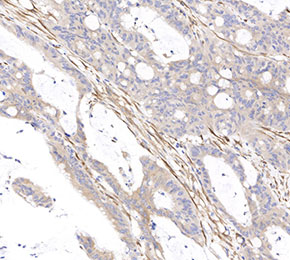 Immunohistochemical analysis of formalin fixed paraffin embedded human colon tissue with F2230 at 1:25000 dilution.