
|
受注:045-509-1970 |
技術サポート:tech@selleck.co.jp 平日9:00〜18:00 1営業日以内にご連絡を差し上げます |
生物学的記述
| Specificity | Orexin A Antibody (Rabbit mAb) [P6H3] detects endogenous levels of total Orexin A peptide. |
|---|---|
| Background | Orexin A, together with orexin B, is one of two neuropeptides derived by proteolytic cleavage from a single prepro-orexin precursor, synthesized exclusively by neurons localized within the lateral and posterior hypothalamus. Both peptides bind and activate two closely related G protein-coupled receptors, OX1R and OX2R, with orexin A engaging both receptor subtypes with comparable affinity, while orexin B shows selectivity favoring OX2R, giving the two-peptide, two-receptor system built-in signaling diversity. Orexin neurons project extensively throughout the central nervous system, including to major nuclei governing sleep-wake regulation, and central administration of orexin A directly stimulates food consumption, linking hypothalamic orexin signaling to acute feeding behavior. Prepro-orexin mRNA expression rises during fasting, positioning the orexin system as a feedback mediator that couples nutritional state to feeding drive rather than acting as a constitutively active signal. Orexin neurons are directly regulated by circulating glucose, leptin, and ghrelin, and this responsiveness allows the orexin system to adjust arousal output according to whole-body energy balance, increasing wakefulness during states of energy deficit such as fasting. OX2R signaling is the primary driver of sleep-wake state stabilization, with receptor activity in the tuberomammillary nucleus and basal forebrain regions maintaining wakefulness, while OX1R signaling contributes additionally to reward-related and motivated behaviors through projections to mesolimbic circuits. Genetic loss of orexin neurons or disruption of OX2R signaling produces a phenotype of poor wakefulness maintenance and cataplexy that closely mirrors human narcolepsy, and this loss-of-function relationship has directly informed the development of OX2R-selective agonists as a therapeutic strategy targeting the underlying neurochemical deficit in narcolepsy type 1, distinct from orexin receptor antagonists developed for insomnia by blocking the same wake-promoting pathway. |
使用情報
| Application | IHC, ELISA | Dilution |
|
||
|---|---|---|---|---|---|
| Reactivity | Mouse, Rat | ||||
| Source | Rabbit Monoclonal Antibody | MW | 13 kDa | ||
| Storage Buffer | PBS, pH 7.2+50% Glycerol+0.05% BSA+0.01% NaN3 | Storage (from the date of receipt) |
-20°C (avoid freeze-thaw cycles), 2 years | ||
| IHC |
Experimental Protocol:
Deparaffinization/Rehydration
1. Deparaffinize/hydrate sections:
2. Incubate sections in three washes of xylene for 5 min each.
3. Incubate sections in two washes of 100% ethanol for 10 min each.
4. Incubate sections in two washes of 95% ethanol for 10 min each.
5. Wash sections two times in dH2O for 5 min each.
6.Antigen retrieval: For Citrate: Heat slides in a microwave submersed in 1X citrate unmasking solution until boiling is initiated; continue with 10 min at a sub-boiling temperature (95°-98°C). Cool slides on bench top for 30 min.
Staining
1. Wash sections in dH2O three times for 5 min each.
2. Incubate sections in 3% hydrogen peroxide for 10 min.
3. Wash sections in dH2O two times for 5 min each.
4. Wash sections in wash buffer for 5 min.
5. Block each section with 100–400 µl of blocking solution for 1 hr at room temperature.
6. Remove blocking solution and add 100–400 µl primary antibody diluent in to each section. Incubate overnight at 4°C.
7. Remove antibody solution and wash sections with wash buffer three times for 5 min each.
8. Cover section with 1–3 drops HRPas needed. Incubate in a humidified chamber for 30 min at room temperature.
9. Wash sections three times with wash buffer for 5 min each.
10. Add DAB Chromogen Concentrate to DAB Diluent and mix well before use.
11. Apply 100–400 µl DAB to each section and monitor closely. 1–10 min generally provides an acceptable staining intensity.
12. Immerse slides in dH2O.
13. If desired, counterstain sections with hematoxylin.
14. Wash sections in dH2O two times for 5 min each.
15. Dehydrate sections: Incubate sections in 95% ethanol two times for 10 sec each; Repeat in 100% ethanol, incubating sections two times for 10 sec each; Repeat in xylene, incubating sections two times for 10 sec each.
16. Mount sections with coverslips and mounting medium.
|
References
|
Application Data
IHC
Validated by Selleck
-
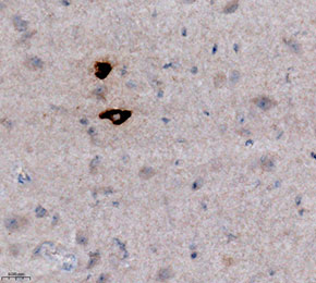 Immunohistochemical analysis of formalin fixed paraffin embedded mouse hypothalamus tissue with F8328 at 1:1000 dilution.