|
受注:045-509-1970 |
技術サポート:tech@selleck.co.jp 平日9:00〜18:00 1営業日以内にご連絡を差し上げます |
生物学的記述
| Specificity | p57 Kip2 Antibody [B21E12] detects endogenous levels of total p57 Kip2 protein. |
|---|---|
| Background | p57 Kip2 (CDKN1C) belongs to the Cip/Kip family of cyclin‑dependent kinase inhibitors together with p21 and p27, and acts as a potent negative regulator of cell proliferation that is encoded by a maternally expressed imprinted gene at chromosome 11p15.5 within a growth‑control domain frequently altered in imprinting disorders and cancer. The protein contains an N‑terminal CDK inhibitory domain that is conserved across Cip/Kip members and binds G1 cyclin–CDK complexes including cyclin D/CDK4–6 and cyclin E/CDK2, enforcing G1 arrest, followed by a unique central proline‑rich region that interacts with the LIM‑kinase LIMK1 and a C‑terminal QT domain that associates with and inhibits JNK/SAPK, creating a modular architecture that couples classical cell‑cycle inhibition with direct control of cytoskeletal dynamics and stress‑activated kinase signaling. The LIMK1‑binding central region localizes LIMK1 in the nucleus and modifies cofilin phosphorylation and actin filament organization, linking p57 Kip2 to regulation of cell shape, migration, and neuronal process outgrowth in addition to its role in proliferation control. The QT domain binds JNK/SAPK and dampens its kinase activity, influencing apoptosis and differentiation programs downstream of stress and developmental cues, while p57 Kip2 also modulates genome expression more broadly through reported interactions with transcriptional regulators and chromatin‑associated complexes, providing a route by which CDK inhibition is integrated with lineage‑specific transcriptional outputs. Expression is tightly developmentally regulated and far more restricted than other Cip/Kip proteins, with high levels in placenta, developing brain, muscle, and hematopoietic compartments, and induction by pathways such as TGF‑β/Smad in hematopoietic stem cells where p57 Kip2 enforces quiescence, contrasted with TGF‑β‑driven degradation in osteoblasts, illustrating context‑dependent integration of extracellular signals. Protein stability is controlled by phosphorylation‑dependent ubiquitination: phosphorylation on threonine 310 promotes recognition by the SCF–Skp2 E3 ubiquitin ligase complex, targeting p57 Kip2 for proteasomal degradation in a mechanism analogous to Thr187‑dependent turnover of p27, and placing p57 Kip2 under direct control of mitogenic CDK–Skp2 circuits. In addition to its central function in restraining G1–S transition, p57 Kip2 plays critical roles at early steps of cell and tissue differentiation, particularly in embryogenesis, neuronal development, and erythropoiesis, where its presence coordinates cell‑cycle exit with acquisition of differentiated phenotypes. The protein influences cell migration through LIMK1‑dependent reorganization of the actin cytoskeleton, and emerging evidence indicates that p57 Kip2 also participates in regulation of mitochondrial apoptotic pathways, with specific phosphorylation patterns modulating its subcellular localization between nucleus and cytoplasm and thereby shifting its impact on survival versus death decisions. CDKN1C is frequently downregulated by genetic, epigenetic, or imprinting alterations in a broad spectrum of epithelial and non‑epithelial malignancies, consistent with its classification as a tumor suppressor, and loss or reduction of p57 Kip2 correlates with enhanced proliferation, invasion, and metastatic behavior, while enforced expression suppresses tumorigenic traits in multiple models. Germline loss‑of‑function mutations and epimutations affecting CDKN1C contribute to Beckwith–Wiedemann syndrome, characterized by somatic overgrowth and elevated childhood tumor risk, whereas distinct gain‑of‑function mutations underlie IMAGe syndrome with severe growth restriction, illustrating that finely tuned p57 Kip2 dosage is required for normal human growth control and that both reduced and excessive activity are pathogenic. |
使用情報
| Application | WB, IHC | Dilution |
|
||||
|---|---|---|---|---|---|---|---|
| Reactivity | Human, Mouse | ||||||
| Source | Mouse Monoclonal Antibody | MW | 32 kDa | ||||
| Storage Buffer | PBS, pH 7.2+50% Glycerol+0.05% BSA+0.01% NaN3 | Storage (from the date of receipt) |
-20°C (avoid freeze-thaw cycles), 2 years | ||||
References
|
Application Data
WB
Validated by Selleck
-
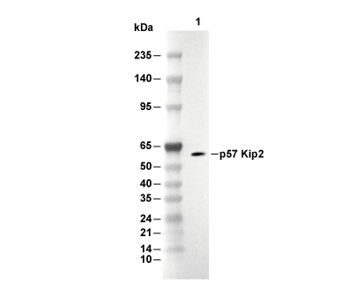 Lane 1: SH-SY5Y