|
受注:045-509-1970 |
技術サポート:tech@selleck.co.jp 平日9:00〜18:00 1営業日以内にご連絡を差し上げます |
生物学的記述
| Specificity | Phospho-FGF Receptor (Tyr653/654) Antibody [F13P7] detects endogenous levels of total FGF Receptor protein only when it is phosphorylated at Tyr653/654. |
|---|---|
| Background | Phospho-FGF Receptor (Tyr653/654) is the principal activation mark of FGFR1-4, driving the transition of these receptors from a low- to high-activity state upon ligand-induced dimerization, a process that underpins FGF-dependent control of cell proliferation, differentiation, migration, and survival. This phosphorylation occurs in the kinase activation loop, precisely between the DFG and APE motifs, and is absolutely required for full catalytic activity and efficient signal propagation. Tyr653 phosphorylation primes the kinase for activity, while subsequent Tyr654 phosphorylation fully stabilizes the active conformation, enabling broad substrate access and robust engagement of downstream signaling partners such as PLCγ, FRS2α, and Crk. These interactions relay signals to key pathways, including MAPK/ERK and PI3K/AKT, regulate gene expression, cytoskeletal dynamics, and cellular metabolism, and determine cell fate decisions during embryogenesis and tissue repair. Phospho-Tyr653/654 also integrates extracellular matrix cues and can be induced in a ligand-independent manner through receptor clustering or oncogenic mutations, contributing to aberrant FGFR signaling. Dephosphorylation by protein tyrosine phosphatases tightly regulates signal duration and amplitude. Mutations affecting Tyr653/654 or the activation loop are causative for skeletal dysplasias such as thanatophoric dysplasia, craniosynostosis syndromes, and dwarfism, by either constitutively activating or inactivating FGFR function. In cancer, persistent phosphorylation at these sites, often due to activating FGFR3 mutations or gene amplifications, drives uncontrolled proliferation, angiogenesis, and therapy resistance. |
使用情報
| Application | WB | Dilution |
|
||
|---|---|---|---|---|---|
| Reactivity | Human, Monkey | ||||
| Source | Mouse Monoclonal Antibody | MW | 120 kDa, 145 kDa | ||
| Storage Buffer | PBS, pH 7.2+50% Glycerol+0.05% BSA+0.01% NaN3 | Storage (from the date of receipt) |
-20°C (avoid freeze-thaw cycles), 2 years | ||
| WB |
Experimental Protocol:
Sample preparation
1. Tissue: Lyse the tissue sample by adding an appropriate volume of ice-cold RIPA/NP-40 Lysis Buffer (containing Protease Inhibitor Cocktail, Phosphatase Inhibitor Cocktail),and homogenize the tissue at a low temperature. 2. Adherent cell: Aspirate the culture medium and wash the cells with ice-cold PBS twice. Lyse the cells by adding an appropriate volume of RIPA/NP-40 Lysis Buffer (containing Protease Inhibitor Cocktail, Phosphatase Inhibitor Cocktail) and put the sample on ice for 5 min. 3. Suspension cell: Transfer the culture medium to a pre-cooled centrifuge tube. Centrifuge and aspirate the supernatant. Wash the cells with ice-cold PBS twice. Lyse the cells by adding an appropriate volume of RIPA/NP-40 Lysis Buffer (containing Protease Inhibitor Cocktail, Phosphatase Inhibitor Cocktail) and put the sample on ice for 5 min. 4. Place the lysate into a pre-cooled microcentrifuge tube. Centrifuge at 4°C for 15 min. Collect the supernatant;
5. Remove a small volume of lysate to determine the protein concentration;
6. Combine the lysate with protein loading buffer. Boil 20 µL sample under 95-100°C for 5 min. Centrifuge for 5 min after cool down on ice.
Electrophoretic separation
1. According to the concentration of extracted protein, load appropriate amount of protein sample and marker onto SDS-PAGE gels for electrophoresis. Recommended separating gel (lower gel) concentration: 5%. Reference Table for Selecting SDS-PAGE Separation Gel Concentrations 2. Power up 80V for 30 minutes. Then the power supply is adjusted (110 V~150 V), the Marker is observed, and the electrophoresis can be stopped when the indicator band of the predyed protein Marker where the protein is located is properly separated. (Note that the current should not be too large when electrophoresis, too large current (more than 150 mA) will cause the temperature to rise, affecting the result of running glue. If high currents cannot be avoided, an ice bath can be used to cool the bath.)
Transfer membrane
1. Take out the converter, soak the clip and consumables in the pre-cooled converter;
2. Activate PVDF membrane with methanol for 1 min and rinse with transfer buffer;
3. Install it in the order of "black edge of clip - sponge - filter paper - filter paper - glue -PVDF membrane - filter paper - filter paper - sponge - white edge of clip"; 4. The protein was electrotransferred to PVDF membrane. ( 0.45 µm PVDF membrane is recommended ) Reference Table for Selecting PVDF Membrane Pore Size Specifications Recommended conditions for wet transfer: 200 mA, 120 min. ( Note that the transfer conditions can be adjusted according to the protein size. For high-molecular-weight proteins, a higher current and longer transfer time are recommended. However, ensure that the transfer tank remains at a low temperature to prevent gel melting.)
Block
1. After electrotransfer, wash the film with TBST at room temperature for 5 minutes;
2. Incubate the film in the blocking solution ( recommending 5% BSA solution)
for 1 hour at room temperature;
3. Wash the film with TBST for 3 times, 5 minutes each time.
Antibody incubation
1. Use 5% skim milk powder to prepare the primary antibody working liquid (recommended dilution ratio for primary antibody 1:1000), gently shake and incubate with the film at 4°C overnight; 2. Wash the film with TBST 3 times, 5 minutes each time;
3. Add the secondary antibody to the blocking solution and incubate with the film gently at room temperature for 1 hour;
4. After incubation, wash the film with TBST 3 times for 5 minutes each time.
Antibody staining
1. Add the prepared ECL luminescent substrate (or select other color developing substrate according to the second antibody) and mix evenly;
2. Incubate with the film for 1 minute, remove excess substrate (keep the film moist), wrap with plastic film, and expose in the imaging system.
|
References
|
Application Data
WB
Validated by Selleck
-
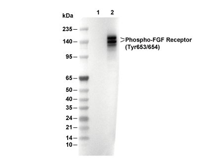 Lane 1: A-204, Lane 2: A-204 (bFGF-treated)