|
受注:045-509-1970 |
技術サポート:tech@selleck.co.jp 平日9:00〜18:00 1営業日以内にご連絡を差し上げます |
生物学的記述
| Specificity | Phospho-RIP (Ser166) Antibody [J17A23] detects endogenous levels of total RIP protein only when it is phosphorylated at Ser166. |
|---|---|
| Background | Phospho-RIP (Ser166), specifically receptor-interacting serine/threonine kinase 1 (RIPK1), is a central adaptor kinase in the RIP kinase family regulating TNFR1-mediated inflammation and cell death, featuring an N-terminal kinase domain (RIP-homologous, auto-inhibitory in basal state), intermediate domain with RHIM motifs for necroptosis, and C-terminal death domain (DD) recruiting TRADD/FADD/caspase-8, where Ser166 autophosphorylation within the activation loop (T-loop) occurs post-TAK1 priming to fully activate catalytic competence. Ser166 phosphorylation (consensus for PIKK kinases) induces T-loop repositioning , DFG motif ordering, C-helix docking, and enhanced ATP/substrate access, enabling trans-autophosphorylation at secondary sites (Ser161/Thr169) while licensing RHIM-dependent RIPK3 recruitment into necrosome (complex IIb). It licenses RIPK1 kinase activity to drive caspase-8/FADD-dependent apoptosis (complex IIa) or MLKL phosphorylation/oligomerization for necroptosis under caspase inhibition (e.g., TSZ/LZ/PZ stimuli), with S166A mutants exhibiting reduced necroptosis/apoptosis, impaired necrosome assembly, and failure to phosphorylate RIPK3/MLKL despite intact kinase domain. phospho-Ser166 sustains inflammatory signaling (NF-κB via IKK/NEMO recruitment, cytokine production) while tipping the balance toward death under unresolved stress; essential for pathogenesis as S166A knock-in mice resist RIPK1-dependent skin inflammation, intestinal damage, hepatitis, and systemic shock. Disease relevance includes inflammatory pathologies such as psoriasis, IBD, and NASH, where it serves as a biomarker for kinase activation; neurodegeneration, with neuroprotection achieved via Nec-1 inhibition; and cancer, where evasion occurs through survival bias. |
使用情報
| Application | WB | Dilution |
|
||
|---|---|---|---|---|---|
| Reactivity | Human | ||||
| Source | Rabbit Monoclonal Antibody | MW | 78-82 kDa | ||
| Storage Buffer | PBS, pH 7.2+50% Glycerol+0.05% BSA+0.01% NaN3 | Storage (from the date of receipt) |
-20°C (avoid freeze-thaw cycles), 2 years | ||
| WB |
Experimental Protocol:
Sample preparation
1. Tissue: Lyse the tissue sample by adding an appropriate volume of ice-cold RIPA/NP-40 Lysis Buffer (containing Protease Inhibitor Cocktail, Phosphatase Inhibitor Cocktail),and homogenize the tissue at a low temperature. 2. Adherent cell: Aspirate the culture medium and wash the cells with ice-cold PBS twice. Lyse the cells by adding an appropriate volume of RIPA/NP-40 Lysis Buffer (containing Protease Inhibitor Cocktail, Phosphatase Inhibitor Cocktail) and put the sample on ice for 5 min. 3. Suspension cell: Transfer the culture medium to a pre-cooled centrifuge tube. Centrifuge and aspirate the supernatant. Wash the cells with ice-cold PBS twice. Lyse the cells by adding an appropriate volume of RIPA/NP-40 Lysis Buffer (containing Protease Inhibitor Cocktail, Phosphatase Inhibitor Cocktail) and put the sample on ice for 5 min. 4. Place the lysate into a pre-cooled microcentrifuge tube. Centrifuge at 4°C for 15 min. Collect the supernatant;
5. Remove a small volume of lysate to determine the protein concentration;
6. Combine the lysate with protein loading buffer. Boil 20 µL sample under 95-100°C for 5 min. Centrifuge for 5 min after cool down on ice.
2. Power up 80V for 30 minutes. Then the power supply is adjusted (110 V~150 V), the Marker is observed, and the electrophoresis can be stopped when the indicator band of the predyed protein Marker where the protein is located is properly separated. (Note that the current should not be too large when electrophoresis, too large current (more than 150 mA) will cause the temperature to rise, affecting the result of running glue. If high currents cannot be avoided, an ice bath can be used to cool the bath.)
Transfer membrane
1. Take out the converter, soak the clip and consumables in the pre-cooled converter;
2. Activate PVDF membrane with methanol for 1 min and rinse with transfer buffer;
3. Install it in the order of "black edge of clip - sponge - filter paper - filter paper - glue -PVDF membrane - filter paper - filter paper - sponge - white edge of clip"; 4. The protein was electrotransferred to PVDF membrane. ( 0.45 µm PVDF membrane is recommended ) Reference Table for Selecting PVDF Membrane Pore Size Specifications Recommended conditions for wet transfer: 200 mA, 120 min. ( Note that the transfer conditions can be adjusted according to the protein size. For high-molecular-weight proteins, a higher current and longer transfer time are recommended. However, ensure that the transfer tank remains at a low temperature to prevent gel melting.)
Block
1. After electrotransfer, wash the film with TBST at room temperature for 5 minutes;
2. Incubate the film in the blocking solution ( recommending 5% BSA solution)
for 1 hour at room temperature;
3. Wash the film with TBST for 3 times, 5 minutes each time.
Antibody incubation
1. Use 5% skim milk powder to prepare the primary antibody working liquid (recommended dilution ratio for primary antibody 1:1000), gently shake and incubate with the film at 4°C overnight; 2. Wash the film with TBST 3 times, 5 minutes each time;
3. Add the secondary antibody to the blocking solution and incubate with the film gently at room temperature for 1 hour;
4. After incubation, wash the film with TBST 3 times for 5 minutes each time.
Antibody staining
1. Add the prepared ECL luminescent substrate (or select other color developing substrate according to the second antibody) and mix evenly;
2. Incubate with the film for 1 minute, remove excess substrate (keep the film moist), wrap with plastic film, and expose in the imaging system.
|
References
|
Application Data
WB
Validated by Selleck
-
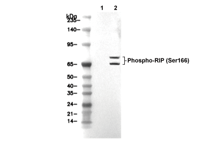 Lane 1: HT-29, Lane 2: HT-29 (Z-VAD, 20 μM, added 30 min prior to other cpds; hTNF-α, 20 ng/ml, 7 h; SM-164, 100 nM, 7 h)