|
受注:045-509-1970 |
技術サポート:tech@selleck.co.jp 平日9:00〜18:00 1営業日以内にご連絡を差し上げます |
生物学的記述
| Specificity | Phospho-VEGF Receptor 2 (Tyr1059) Antibody [J6L3] detects endogenous levels of total VEGF receptor 2 protein only when it is phosphorylated at Tyr1059. |
|---|---|
| Background | Phospho-VEGF Receptor 2 (Tyr1059) refers to the activated form of vascular endothelial growth factor receptor 2 (VEGFR2) in which tyrosine 1059, located within the activation loop of the kinase domain, has been phosphorylated. VEGFR2 is a class III receptor tyrosine kinase and serves as the principal mediator of vascular development, angiogenesis, and vascular permeability. VEGFR2 is composed of an extracellular region containing seven immunoglobulin-like motifs, a single transmembrane segment, and a cytoplasmic tail with a split tyrosine kinase domain. Upon binding of VEGF-A to its extracellular domain, VEGFR2 undergoes dimerization and autophosphorylation at several cytoplasmic tyrosine residues, with phosphorylation at Tyr1059 being a pivotal event that enhances the catalytic activity of the kinase domain. This phosphorylation triggers the engagement of multiple downstream signaling cascades, including the PLCγ-PKC-MAPK and PI3K/Akt pathways. Through these pathways, Phospho-VEGFR2 (Tyr1059) regulates essential endothelial cell behaviors such as survival, proliferation, migration, and vascular permeability. The activation of VEGFR2 at Tyr1059 is a central driver of angiogenesis, enabling the formation of new blood vessels during embryonic development, wound healing, and tissue repair. However, persistent or aberrant activation of Phospho-VEGFR2 (Tyr1059) is frequently observed in pathological angiogenesis, such as in solid tumors and inflammatory diseases, where it contributes to uncontrolled vessel growth and increased vascular leakiness. Phospho-Tyr1059 VEGFR2 serves as a key biomarker for active VEGFR2 signaling and is a prime therapeutic target. Inhibitors targeting this phosphorylation event are used to suppress aberrant angiogenesis in cancer and ocular diseases. |
使用情報
| Application | WB | Dilution |
|
||
|---|---|---|---|---|---|
| Reactivity | Human, Mouse | ||||
| Source | Rabbit Monoclonal Antibody | MW | 230 kDa | ||
| Storage Buffer | PBS, pH 7.2+50% Glycerol+0.05% BSA+0.01% NaN3 | Storage (from the date of receipt) |
-20°C (avoid freeze-thaw cycles), 2 years | ||
| WB |
Experimental Protocol:
Sample preparation
1. Tissue: Lyse the tissue sample by adding an appropriate volume of ice-cold RIPA/NP-40 Lysis Buffer (containing Protease Inhibitor Cocktail, Phosphatase Inhibitor Cocktail),and homogenize the tissue at a low temperature. 2. Adherent cell: Aspirate the culture medium and wash the cells with ice-cold PBS twice. Lyse the cells by adding an appropriate volume of RIPA/NP-40 Lysis Buffer (containing Protease Inhibitor Cocktail, Phosphatase Inhibitor Cocktail) and put the sample on ice for 5 min. 3. Suspension cell: Transfer the culture medium to a pre-cooled centrifuge tube. Centrifuge and aspirate the supernatant. Wash the cells with ice-cold PBS twice. Lyse the cells by adding an appropriate volume of RIPA/NP-40 Lysis Buffer (containing Protease Inhibitor Cocktail, Phosphatase Inhibitor Cocktail) and put the sample on ice for 5 min. 4. Place the lysate into a pre-cooled microcentrifuge tube. Centrifuge at 4°C for 15 min. Collect the supernatant;
5. Remove a small volume of lysate to determine the protein concentration;
6. Combine the lysate with protein loading buffer. Boil 20 µL sample under 95-100°C for 5 min. Centrifuge for 5 min after cool down on ice.
2. Power up 80V for 30 minutes. Then the power supply is adjusted (110 V~150 V), the Marker is observed, and the electrophoresis can be stopped when the indicator band of the predyed protein Marker where the protein is located is properly separated. (Note that the current should not be too large when electrophoresis, too large current (more than 150 mA) will cause the temperature to rise, affecting the result of running glue. If high currents cannot be avoided, an ice bath can be used to cool the bath.)
Transfer membrane
1. Take out the converter, soak the clip and consumables in the pre-cooled converter;
2. Activate PVDF membrane with methanol for 1 min and rinse with transfer buffer;
3. Install it in the order of "black edge of clip - sponge - filter paper - filter paper - glue -PVDF membrane - filter paper - filter paper - sponge - white edge of clip"; 4. The protein was electrotransferred to PVDF membrane. ( 0.45 µm PVDF membrane is recommended ) Reference Table for Selecting PVDF Membrane Pore Size Specifications Recommended conditions for wet transfer: 250 mA, 180 min. ( Note that the transfer conditions can be adjusted according to the protein size. For high-molecular-weight proteins, a higher current and longer transfer time are recommended. However, ensure that the transfer tank remains at a low temperature to prevent gel melting.)
Block
1. After electrotransfer, wash the film with TBST at room temperature for 5 minutes;
2. Incubate the film in the blocking solution ( recommending 5% BSA solution)
for 1 hour at room temperature;
3. Wash the film with TBST for 3 times, 5 minutes each time.
Antibody incubation
1. Use 5% skim milk powder to prepare the primary antibody working liquid (recommended dilution ratio for primary antibody 1:1000), gently shake and incubate with the film at 4°C overnight; 2. Wash the film with TBST 3 times, 5 minutes each time;
3. Add the secondary antibody to the blocking solution and incubate with the film gently at room temperature for 1 hour;
4. After incubation, wash the film with TBST 3 times for 5 minutes each time.
Antibody staining
1. Add the prepared ECL luminescent substrate (or select other color developing substrate according to the second antibody) and mix evenly;
2. Incubate with the film for 1 minute, remove excess substrate (keep the film moist), wrap with plastic film, and expose in the imaging system.
|
References
|
Application Data
WB
Validated by Selleck
-
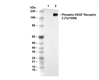 Lane 1: HT-29, Lane 2: HT-29 (SCF-1 treated)