
|
受注:045-509-1970 |
技術サポート:tech@selleck.co.jp 平日9:00〜18:00 1営業日以内にご連絡を差し上げます |
生物学的記述
| Specificity | PSRC2 Antibody (Rabbit mAb) [L8L24] detects endogenous levels of total PSRC2 protein. |
|---|---|
| Background | PSRC2, now catalogued under the official gene symbol ZFC3H1 and carrying the earlier aliases CCDC131, CSRC2, and KIAA0546, encodes a large zinc-knuckle-containing protein that functions as a central adaptor within the mammalian nuclear RNA exosome surveillance network rather than as a classical DNA-binding transcription factor despite its zinc finger nomenclature. The protein carries an N-terminal zinc-knuckle motif alongside an extended, largely coiled-coil scaffold architecture that mediates protein-protein assembly rather than catalytic activity, acting as a structural bridge within multi-subunit RNA decay machinery rather than as an enzymatic subunit itself. ZFC3H1 pairs with the RNA helicase MTR4 to form a tight core heterodimer that constitutes the poly(A) tail exosome targeting connection, commonly termed PAXT, a pathway distinct from and functionally parallel to the trimeric nuclear exosome targeting complex built from MTR4, ZCCHC8, and RBM7. Where the nuclear exosome targeting complex recognises short, early, largely unprocessed RNA species such as promoter-upstream transcripts and enhancer RNAs, the PAXT connection is directed toward longer, more extensively polyadenylated and processed nuclear transcripts, achieving this specificity through an RNA-dependent contact between the MTR4-ZFC3H1 dimer and the nuclear poly(A)-binding protein PABPN1. Depletion of ZFC3H1 or PABPN1 leads to accumulation of a shared cohort of substrate RNAs encompassing both coding and non-coding transcripts, confirming that the two factors act within a single decay branch rather than independently. ZFC3H1 drives the physical sequestration of polyadenylated exosome substrates into discrete nuclear foci that are spatially distinct from Cajal bodies, nuclear speckles, paraspeckles, and nucleoli, and this retention activity is itself required for foci formation. When ZFC3H1 is lost, transcripts that would otherwise be marked for nuclear degradation are instead exported to the cytoplasm through a pathway dependent on the mRNA export factor AlyREF, revealing that PAXT-mediated nuclear retention normally operates in direct competition with the nucleocytoplasmic export machinery to prevent premature transcript escape. This dual capacity for exosome recruitment and physical nuclear tethering determines which processed RNA species are cleared within the nucleus versus permitted onward passage to the cytoplasm, directly shaping noncoding RNA turnover, transcriptional noise clearance, and the fidelity of nuclear RNA quality control. |
使用情報
| Application | WB | Dilution |
|
||
|---|---|---|---|---|---|
| Reactivity | Mouse, Rat, Human | ||||
| Source | Rabbit Monoclonal Antibody | MW | 226 kDa | ||
| Storage Buffer | PBS, pH 7.2+50% Glycerol+0.05% BSA+0.01% NaN3 | Storage (from the date of receipt) |
-20°C (avoid freeze-thaw cycles), 2 years | ||
| WB |
Experimental Protocol:
Sample preparation
1. Tissue: Lyse the tissue sample by adding an appropriate volume of ice-cold RIPA/Nuclear Lysis Buffer (containing Protease Inhibitor Cocktail),and homogenize the tissue at a low temperature or lyse it by sonication on ice, then incubate on ice for 30 minutes. 2. Adherent cell: Aspirate the culture medium and wash the cells with ice-cold PBS twice. Lyse the cells by adding an appropriate volume of RIPA/Nuclear Lysis Buffer (containing Protease Inhibitor Cocktail) , sonicate to lyse the cells, and incubate on ice for 30 minutes. 3. Suspension cell: Transfer the culture medium to a pre-cooled centrifuge tube. Centrifuge and aspirate the supernatant. Wash the cells with ice-cold PBS twice. Lyse the cells by adding an appropriate volume of RIPA/Nuclear Lysis Buffer (containing Protease Inhibitor Cocktail) , sonicate to lyse the cells, and incubate on ice for 30 minutes. 4. Place the lysate into a pre-cooled microcentrifuge tube. Centrifuge at 4°C for 15 min. Collect the supernatant;
5. Remove a small volume of lysate to determine the protein concentration;
6. Combine the lysate with protein loading buffer. Boil 20 µL sample under 95-100°C for 5 min. Centrifuge for 5 min after cool down on ice.
Electrophoretic separation
1. According to the concentration of extracted protein, load appropriate amount of protein sample and marker onto SDS-PAGE gels for electrophoresis. Recommended separating gel (lower gel) concentration: 5%. Reference Table for Selecting SDS-PAGE Separation Gel Concentrations 2. Power up 80V for 30 minutes. Then the power supply is adjusted (110 V~150 V), the Marker is observed, and the electrophoresis can be stopped when the indicator band of the predyed protein Marker where the protein is located is properly separated. (Note that the current should not be too large when electrophoresis, too large current (more than 150 mA) will cause the temperature to rise, affecting the result of running glue. If high currents cannot be avoided, an ice bath can be used to cool the bath.)
Transfer membrane
1. Take out the converter, soak the clip and consumables in the pre-cooled converter;
2. Activate PVDF membrane with methanol for 1 min and rinse with transfer buffer;
3. Install it in the order of "black edge of clip - sponge - filter paper - filter paper - glue -PVDF membrane - filter paper - filter paper - sponge - white edge of clip"; 4. The protein was electrotransferred to PVDF membrane. ( 0.45 µm PVDF membrane is recommended ) Reference Table for Selecting PVDF Membrane Pore Size Specifications Recommended conditions for wet transfer: 250 mA, 180 min. ( Note that the transfer conditions can be adjusted according to the protein size. For high-molecular-weight proteins, a higher current and longer transfer time are recommended. However, ensure that the transfer tank remains at a low temperature to prevent gel melting.)
Block
1. After electrotransfer, wash the film with TBST at room temperature for 5 minutes;
2. Incubate the film in the blocking solution for 1 hour at room temperature;
3. Wash the film with TBST for 3 times, 5 minutes each time.
Antibody incubation
1. Use primary antibody dilution buffer to prepare the primary antibody working liquid (recommended dilution ratio for primary antibody 1:1000), gently shake and incubate with the film at 4°C overnight; 2. Wash the film with TBST 3 times, 5 minutes each time;
3. Add the secondary antibody to the blocking solution and incubate with the film gently at room temperature for 1 hour;
4. After incubation, wash the film with TBST 3 times for 5 minutes each time.
Antibody staining
1. Add the prepared ECL luminescent substrate (or select other color developing substrate according to the second antibody) and mix evenly;
2. Incubate with the film for 1 minute, remove excess substrate (keep the film moist), wrap with plastic film, and expose in the imaging system.
|
References
|
Application Data
WB
Validated by Selleck
-
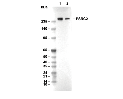 Lane 1: HeLa, Lane 2: 293T