|
受注:045-509-1970 |
技術サポート:tech@selleck.co.jp 平日9:00〜18:00 1営業日以内にご連絡を差し上げます |
生物学的記述
| Specificity | SATB2 Antibody [C16D1] detects endogenous levels of total SATB2 protein. |
|---|---|
| Background | SATB2 (special AT‑rich sequence‑binding protein 2) is a MAR‑binding nuclear protein of the SATB family that functions as a higher‑order chromatin organizer and transcriptional regulator, integrating long‑range chromatin architecture with gene expression programs governing craniofacial and skeletal development, neocortical projection neuron identity, hematopoietic differentiation, and tumorigenesis. The protein contains two CUT domains and a homeodomain embedded in a low‑complexity N‑terminal region that tether SATB2 to AT‑rich matrix attachment region elements and recruit chromatin modifiers and transcriptional co‑regulators, allowing SATB2 to assemble regulatory hubs that loop distant genomic elements into functional domains and either activate or repress target gene clusters depending on associated cofactors. In osteoblast‑lineage cells of branchial arches and developing bone, SATB2 promotes osteogenic differentiation and craniofacial patterning by repressing posterior Hoxa2 expression and cooperating with osteogenic transcription factors Runx2 and ATF4 to induce bone matrix genes, and its dosage critically influences craniofacial morphogenesis and bone regeneration potential, establishing SATB2 as an osteoinductive factor with translational relevance for bone defect repair. In the developing cerebral cortex, SATB2 is expressed in callosal and subsets of subcerebral projection neurons, where it binds MARs and regulatory regions near Ctip2/Bcl11b and other corticofugal determinants to repress subcortical identity programs and promote callosal projection neuron fate, thereby directing axon targeting across the corpus callosum rather than down corticospinal tracts; conditional deletion reveals additional roles in deep‑layer subcerebral projection neurons, where early transient SATB2 expression is required for corticospinal tract formation, demonstrating layer‑ and time‑dependent control of projection neuron subclass specification and axon pathfinding. Genome‑wide mapping of SATB2 targets in developing cortex identifies more than a thousand downstream effector genes organized into temporally distinct clusters, including axon guidance receptors and ligands such as Plxnd1, Ntng1, Efnb2, Ephb1, Plxna2, and Epha3, as well as synapse‑related genes and the transcription factor Mef2c, placing SATB2 at the apex of regulatory networks that coordinate axonogenesis, synaptogenesis, and synaptic plasticity and linking SATB2 dosage to cognitive and psychiatric phenotypes through enrichment of SATB2‑regulated genes among neurodevelopmental disorder loci. In the adult cortex and hippocampus, SATB2 persists in pyramidal neurons across layers, where it switches interaction partners from predominantly repressive complexes in development to chromatin‑structuring and transcription‑modulating networks in maturity, and SATB2‑interacting gene sets are highly constrained in humans, with rare disruptive variants causing severe cognitive disorders and common variants contributing to general cognitive ability, emphasizing the importance of SATB2‑centered chromatin hubs in human cognition. |
使用情報
| Application | WB, IP, IHC | Dilution |
|
||||||
|---|---|---|---|---|---|---|---|---|---|
| Reactivity | Human | ||||||||
| Source | Rabbit Monoclonal Antibody | MW | 83 kDa | ||||||
| Storage Buffer | PBS, pH 7.2+50% Glycerol+0.05% BSA+0.01% NaN3 | Storage (from the date of receipt) |
-20°C (avoid freeze-thaw cycles), 2 years | ||||||
| WB |
Experimental Protocol:
Sample preparation
1. Tissue: Lyse the tissue sample by adding an appropriate volume of ice-cold RIPA/Nuclear Lysis Buffer (containing Protease Inhibitor Cocktail),and homogenize the tissue at a low temperature or lyse it by sonication on ice, then incubate on ice for 30 minutes. 2. Adherent cell: Aspirate the culture medium and wash the cells with ice-cold PBS twice. Lyse the cells by adding an appropriate volume of RIPA/Nuclear Lysis Buffer (containing Protease Inhibitor Cocktail) , sonicate to lyse the cells, and incubate on ice for 30 minutes. 3. Suspension cell: Transfer the culture medium to a pre-cooled centrifuge tube. Centrifuge and aspirate the supernatant. Wash the cells with ice-cold PBS twice. Lyse the cells by adding an appropriate volume of RIPA/Nuclear Lysis Buffer (containing Protease Inhibitor Cocktail) , sonicate to lyse the cells, and incubate on ice for 30 minutes. 4. Place the lysate into a pre-cooled microcentrifuge tube. Centrifuge at 4°C for 15 min. Collect the supernatant;
5. Remove a small volume of lysate to determine the protein concentration;
6. Combine the lysate with protein loading buffer. Boil 20 µL sample under 95-100°C for 5 min. Centrifuge for 5 min after cool down on ice.
Electrophoretic separation
1. According to the concentration of extracted protein, load appropriate amount of protein sample and marker onto SDS-PAGE gels for electrophoresis. Recommended separating gel (lower gel) concentration: 10%. Reference Table for Selecting SDS-PAGE Separation Gel Concentrations 2. Power up 80V for 30 minutes. Then the power supply is adjusted (110 V~150 V), the Marker is observed, and the electrophoresis can be stopped when the indicator band of the predyed protein Marker where the protein is located is properly separated. (Note that the current should not be too large when electrophoresis, too large current (more than 150 mA) will cause the temperature to rise, affecting the result of running glue. If high currents cannot be avoided, an ice bath can be used to cool the bath.)
Transfer membrane
1. Take out the converter, soak the clip and consumables in the pre-cooled converter;
2. Activate PVDF membrane with methanol for 1 min and rinse with transfer buffer;
3. Install it in the order of "black edge of clip - sponge - filter paper - filter paper - glue -PVDF membrane - filter paper - filter paper - sponge - white edge of clip"; 4. The protein was electrotransferred to PVDF membrane. ( 0.45 µm PVDF membrane is recommended ) Reference Table for Selecting PVDF Membrane Pore Size Specifications Recommended conditions for wet transfer: 200 mA, 120 min. ( Note that the transfer conditions can be adjusted according to the protein size. For high-molecular-weight proteins, a higher current and longer transfer time are recommended. However, ensure that the transfer tank remains at a low temperature to prevent gel melting.)
Block
1. After electrotransfer, wash the film with TBST at room temperature for 5 minutes;
2. Incubate the film in the blocking solution for 1 hour at room temperature;
3. Wash the film with TBST for 3 times, 5 minutes each time.
Antibody incubation
1. Use 5% skim milk powder to prepare the primary antibody working liquid (recommended dilution ratio for primary antibody 1:1000), gently shake and incubate with the film at 4°C overnight; 2. Wash the film with TBST 3 times, 5 minutes each time;
3. Add the secondary antibody to the blocking solution and incubate with the film gently at room temperature for 1 hour;
4. After incubation, wash the film with TBST 3 times for 5 minutes each time.
Antibody staining
1. Add the prepared ECL luminescent substrate (or select other color developing substrate according to the second antibody) and mix evenly;
2. Incubate with the film for 1 minute, remove excess substrate (keep the film moist), wrap with plastic film, and expose in the imaging system.
|
| IHC |
Experimental Protocol:
Deparaffinization/Rehydration
1. Deparaffinize/hydrate sections:
2. Incubate sections in three washes of xylene for 5 min each.
3. Incubate sections in two washes of 100% ethanol for 10 min each.
4. Incubate sections in two washes of 95% ethanol for 10 min each.
5. Wash sections two times in dH2O for 5 min each.
6.Antigen retrieval: For Citrate: Heat slides in a microwave submersed in 1X citrate unmasking solution until boiling is initiated; continue with 10 min at a sub-boiling temperature (95°-98°C). Cool slides on bench top for 30 min.
Staining
1. Wash sections in dH2O three times for 5 min each.
2. Incubate sections in 3% hydrogen peroxide for 10 min.
3. Wash sections in dH2O two times for 5 min each.
4. Wash sections in wash buffer for 5 min.
5. Block each section with 100–400 µl of blocking solution for 1 hr at room temperature.
6. Remove blocking solution and add 100–400 µl primary antibody diluent in to each section. Incubate overnight at 4°C.
7. Remove antibody solution and wash sections with wash buffer three times for 5 min each.
8. Cover section with 1–3 drops HRPas needed. Incubate in a humidified chamber for 30 min at room temperature.
9. Wash sections three times with wash buffer for 5 min each.
10. Add DAB Chromogen Concentrate to DAB Diluent and mix well before use.
11. Apply 100–400 µl DAB to each section and monitor closely. 1–10 min generally provides an acceptable staining intensity.
12. Immerse slides in dH2O.
13. If desired, counterstain sections with hematoxylin.
14. Wash sections in dH2O two times for 5 min each.
15. Dehydrate sections: Incubate sections in 95% ethanol two times for 10 sec each; Repeat in 100% ethanol, incubating sections two times for 10 sec each; Repeat in xylene, incubating sections two times for 10 sec each.
16. Mount sections with coverslips and mounting medium.
|
References
|
Application Data
WB
Validated by Selleck
-
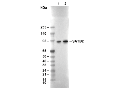 Lane 1: DLD-1, Lane 2: THP-1