|
受注:045-509-1970 |
技術サポート:tech@selleck.co.jp 平日9:00〜18:00 1営業日以内にご連絡を差し上げます |
生物学的記述
| Specificity | Sharpin Antibody [G5P13] detects endogenous levels of total Sharpin protein. |
|---|---|
| Background | Sharpin belongs to the LUBAC family of ubiquitin ligases, alongside HOIP and HOIL-1L, and serves as a key adaptor in linear ubiquitin chain formation. The protein features an N-terminal pleckstrin homology (PH) domain forming a tetrameric superfold that drives dimerization, a central ubiquitin-like (UBL) domain for complex assembly, and a C-terminal NPL4 zinc finger (NZF) domain that binds linear polyubiquitin chains. These domains position Sharpin within the LUBAC to conjugate Met1-linked ubiquitin onto substrates like NEMO and RIPK1 following TNF-α or IL-1 stimulation. Sharpin stabilizes LUBAC through LTM motifs that co-fold into a tethering domain with HOIL-1L, enhancing HOIP's RBR ligase activity for NF-κB pathway activation. This linear ubiquitination amplifies canonical NF-κB signaling, promoting transcription of cytokines, anti-apoptotic genes, and survival factors in immune cells. Sharpin recruits LUBAC to TNFR1 complex I, preventing RIPK1-mediated necroptosis by counterbalancing caspase-8 cleavage. Sharpin deficiency disrupts B-cell development and marginal zone formation through impaired RelA activation. The NZF domain senses ubiquitin chains to amplify signaling feedback loops during inflammation. Loss-of-function mutations in mice trigger chronic proliferative dermatitis with eosinophilic infiltration, ulceration, and runting due to excessive TNFR1-induced apoptosis and inflammation. Sharpin overexpression promotes proliferation, invasion, and p53 degradation via MDM2-mediated polyubiquitination, driving breast and renal cell carcinoma progression. It also enhances PI3K/AKT signaling by targeting PTEN and boosts melanoma growth through PRMT5/SOX10 axis activation. Sharpin mutations link to autoinflammatory syndromes, underscoring its role in balancing immune homeostasis and preventing excessive tissue damage. |
使用情報
| Application | WB, IP | Dilution |
|
||||
|---|---|---|---|---|---|---|---|
| Reactivity | Human, Monkey | ||||||
| Source | Rabbit Monoclonal Antibody | MW | 43 kDa | ||||
| Storage Buffer | PBS, pH 7.2+50% Glycerol+0.05% BSA+0.01% NaN3 | Storage (from the date of receipt) |
-20°C (avoid freeze-thaw cycles), 2 years | ||||
| WB |
Experimental Protocol:
Sample preparation
1. Tissue: Lyse the tissue sample by adding an appropriate volume of ice-cold RIPA/NP-40 Lysis Buffer (containing Protease Inhibitor Cocktail),and homogenize the tissue at a low temperature. 2. Adherent cell: Aspirate the culture medium and wash the cells with ice-cold PBS twice. Lyse the cells by adding an appropriate volume of RIPA/NP-40 Lysis Buffer (containing Protease Inhibitor Cocktail) and put the sample on ice for 5 min. 3. Suspension cell: Transfer the culture medium to a pre-cooled centrifuge tube. Centrifuge and aspirate the supernatant. Wash the cells with ice-cold PBS twice. Lyse the cells by adding an appropriate volume of RIPA/NP-40 Lysis Buffer (containing Protease Inhibitor Cocktail) and put the sample on ice for 5 min. 4. Place the lysate into a pre-cooled microcentrifuge tube. Centrifuge at 4°C for 15 min. Collect the supernatant;
5. Remove a small volume of lysate to determine the protein concentration;
6. Combine the lysate with protein loading buffer. Boil 20 µL sample under 95-100°C for 5 min. Centrifuge for 5 min after cool down on ice.
2. Power up 80V for 30 minutes. Then the power supply is adjusted (110 V~150 V), the Marker is observed, and the electrophoresis can be stopped when the indicator band of the predyed protein Marker where the protein is located is properly separated. (Note that the current should not be too large when electrophoresis, too large current (more than 150 mA) will cause the temperature to rise, affecting the result of running glue. If high currents cannot be avoided, an ice bath can be used to cool the bath.)
Transfer membrane
1. Take out the converter, soak the clip and consumables in the pre-cooled converter;
2. Activate PVDF membrane with methanol for 1 min and rinse with transfer buffer;
3. Install it in the order of "black edge of clip - sponge - filter paper - filter paper - glue -PVDF membrane - filter paper - filter paper - sponge - white edge of clip"; 4. The protein was electrotransferred to PVDF membrane. ( 0.45 µm PVDF membrane is recommended ) Reference Table for Selecting PVDF Membrane Pore Size Specifications Recommended conditions for wet transfer: 200 mA, 120 min. ( Note that the transfer conditions can be adjusted according to the protein size. For high-molecular-weight proteins, a higher current and longer transfer time are recommended. However, ensure that the transfer tank remains at a low temperature to prevent gel melting.)
Block
1. After electrotransfer, wash the film with TBST at room temperature for 5 minutes;
2. Incubate the film in the blocking solution for 1 hour at room temperature;
3. Wash the film with TBST for 3 times, 5 minutes each time.
Antibody incubation
1. Use 5% skim milk powder to prepare the primary antibody working liquid (recommended dilution ratio for primary antibody 1:1000), gently shake and incubate with the film at 4°C overnight; 2. Wash the film with TBST 3 times, 5 minutes each time;
3. Add the secondary antibody to the blocking solution and incubate with the film gently at room temperature for 1 hour;
4. After incubation, wash the film with TBST 3 times for 5 minutes each time.
Antibody staining
1. Add the prepared ECL luminescent substrate (or select other color developing substrate according to the second antibody) and mix evenly;
2. Incubate with the film for 1 minute, remove excess substrate (keep the film moist), wrap with plastic film, and expose in the imaging system. (Exposure time of at least 120s is recommended)
|
References
|
Application Data
WB
Validated by Selleck
-
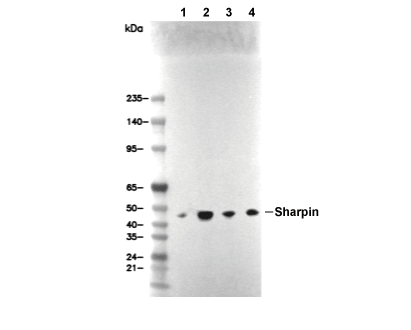 Lane 1: DU145, Lane 2: PC-3, Lane 3: MCF7, Lane 4: T-47D