
|
受注:045-509-1970 |
技術サポート:tech@selleck.co.jp 平日9:00〜18:00 1営業日以内にご連絡を差し上げます |
生物学的記述
| Specificity | SirT4 Antibody (Rabbit mAb) [C24N9] detects endogenous levels of total SirT4 protein. |
|---|---|
| Background | SIRT4 resides within the sirtuin family of NAD+-dependent enzymes, primarily localizing to mitochondria where it exerts regulatory control over metabolic processes distinct from classical deacetylase functions. SIRT4 adopts a conserved catalytic core that accommodates diverse substrates through flexible active site geometry, enabling deacylation, ADP-ribosylation, and lipoamidase activities alongside selective deacetylation. Enzymatic action targets malonyl CoA decarboxylase through deacetylation at specific lysine residues, thereby inhibiting its conversion of malonyl CoA to acetyl CoA and sustaining malonyl CoA pools that allosterically suppress carnitine palmitoyltransferase-1 to curtail fatty acid oxidation while favoring lipogenesis under nutrient-replete states. SIRT4 further modifies glutamine dehydrogenase via ADP-ribosylation, constraining glutaminolysis and TCA cycle anaplerosis to preserve mitochondrial redox balance and limit proliferation signals in response to genotoxic stress. Interaction with the pyruvate dehydrogenase complex occurs through delipoamidation of the E2 subunit dihydrolipoyllysine acetyltransferase, disrupting PDH flux and redirecting carbon toward oxidative phosphorylation or antioxidant defenses during energy crises. SIRT4 coordinates lipid homeostasis in skeletal muscle and adipose tissue by repressing catabolic shifts, while in tumor cells, it couples DNA damage sensing to metabolic arrest via glutamine restriction, activating AMPK-p53 phosphorylation cascades that induce autophagy and suppress Warburg reprogramming. Reduced SIRT4 activity accompanies accelerated tumorigenesis across pancreatic, prostate, and colorectal cancers, where unchecked MCD activation and glutamine flux fuel biomass accumulation and metastatic dissemination. |
使用情報
| Application | WB, IP | Dilution |
|
||||
|---|---|---|---|---|---|---|---|
| Reactivity | Mouse | ||||||
| Source | Rabbit Monoclonal Antibody | MW | 32 kDa | ||||
| Storage Buffer | PBS, pH 7.2+50% Glycerol+0.05% BSA+0.01% NaN3 | Storage (from the date of receipt) |
-20°C (avoid freeze-thaw cycles), 2 years | ||||
| WB |
Experimental Protocol:
Sample preparation
1. Tissue: Lyse the tissue sample by adding an appropriate volume of ice-cold RIPA Lysis Buffer (containing Protease Inhibitor Cocktail),and homogenize the tissue at a low temperature or lyse it by sonication on ice, then incubate on ice for 30 minutes. 2. Adherent cell: Aspirate the culture medium and wash the cells with ice-cold PBS twice. Lyse the cells by adding an appropriate volume of RIPA Lysis Buffer (containing Protease Inhibitor Cocktail), sonicate to lyse the cells, and incubate on ice for 30 minutes. 3. Suspension cell: Transfer the culture medium to a pre-cooled centrifuge tube. Centrifuge and aspirate the supernatant. Wash the cells with ice-cold PBS twice. Lyse the cells by adding an appropriate volume of RIPA Lysis Buffer (containing Protease Inhibitor Cocktail), sonicate to lyse the cells, and incubate on ice for 30 minutes. 4. Place the lysate into a pre-cooled microcentrifuge tube. Centrifuge at 4°C for 15 min. Collect the supernatant;
5. Remove a small volume of lysate to determine the protein concentration;
6. Combine the lysate with protein loading buffer. Boil 20 µL sample under 95-100°C for 5 min. Centrifuge for 5 min after cool down on ice.
Electrophoretic separation
1. According to the concentration of extracted protein, load appropriate amount of protein sample and marker onto SDS-PAGE gels for electrophoresis. Recommended separating gel (lower gel) concentration: 10%. Reference Table for Selecting SDS-PAGE Separation Gel Concentrations 2. Power up 80V for 30 minutes. Then the power supply is adjusted (110 V~150 V), the Marker is observed, and the electrophoresis can be stopped when the indicator band of the predyed protein Marker where the protein is located is properly separated. (Note that the current should not be too large when electrophoresis, too large current (more than 150 mA) will cause the temperature to rise, affecting the result of running glue. If high currents cannot be avoided, an ice bath can be used to cool the bath.)
Transfer membrane
1. Take out the converter, soak the clip and consumables in the pre-cooled converter;
2. Activate PVDF membrane with methanol for 1 min and rinse with transfer buffer;
3. Install it in the order of "black edge of clip - sponge - filter paper - filter paper - glue -PVDF membrane - filter paper - filter paper - sponge - white edge of clip"; 4. The protein was electrotransferred to PVDF membrane. ( 0.45 µm PVDF membrane is recommended ) Reference Table for Selecting PVDF Membrane Pore Size Specifications Recommended conditions for wet transfer: 200 mA, 60 min. ( Note that the transfer conditions can be adjusted according to the protein size. For high-molecular-weight proteins, a higher current and longer transfer time are recommended. However, ensure that the transfer tank remains at a low temperature to prevent gel melting.)
Block
1. After electrotransfer, wash the film with TBST at room temperature for 5 minutes;
2. Incubate the film in the blocking solution for 1 hour at room temperature;
3. Wash the film with TBST for 3 times, 5 minutes each time.
Antibody incubation
1. Use 5% skim milk powder to prepare the primary antibody working liquid (recommended dilution ratio for primary antibody 1:1000), gently shake and incubate with the film at 4°C overnight; 2. Wash the film with TBST 3 times, 5 minutes each time;
3. Add the secondary antibody to the blocking solution and incubate with the film gently at room temperature for 1 hour;
4. After incubation, wash the film with TBST 3 times for 5 minutes each time.
Antibody staining
1. Add the prepared ECL luminescent substrate (or select other color developing substrate according to the second antibody) and mix evenly;
2. Incubate with the film for 1 minute, remove excess substrate (keep the film moist), wrap with plastic film, and expose in the imaging system.
|
References
|
Application Data
WB
Validated by Selleck
-
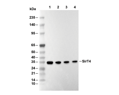 Lane 1: Neuro-2a, Lane 2: C2C12, Lane 3: RAW264.7, Lane 4: NIH/3T3