
|
受注:045-509-1970 |
技術サポート:tech@selleck.co.jp 平日9:00〜18:00 1営業日以内にご連絡を差し上げます |
生物学的記述
| Specificity | TARC/CCL17 Antibody (Rabbit mAb) [P14H22] detects endogenous levels of total TARC/CCL17 protein. |
|---|---|
| Background | CCL17, originally identified as thymus and activation-regulated chemokine, belongs to the CC chemokine subfamily and was first characterized as a T cell-directed chemokine constitutively expressed in the thymus, with inducible expression in peripheral blood mononuclear cells following mitogen stimulation. CCL17 functions as a highly specific biological ligand for the G-protein-coupled receptor CCR4, and engagement requires two distinct binding sites on the chemokine, since antibody blockade of either site independently disrupts CCR4 signaling and abolishes the establishment of localized inflammatory responses. CCR4 is expressed predominantly on Th2 cells alongside multiple other lymphocyte subsets, including skin-homing T cells bearing cutaneous lymphocyte antigen, regulatory T cells, Th17 and Th22 populations, and CCL17 binding drives directional T cell migration together with intracellular calcium mobilization. Downstream of receptor engagement, phospholipase-C-dependent, calcium-independent mechanisms are required for the directional component of T cell migration, distinguishing the motility-driving signal from the calcium flux that accompanies receptor activation, while pertussis toxin sensitivity across multiple cell systems indicates that heterotrimeric G-protein coupling underlies the chemotactic response. CCR4 also engages β-arrestin following ligand binding, and CCL17 and the related ligand CCL22 differ in the degree of receptor internalization and desensitization they produce, a distinction attributed to CCR4 adopting at least two structurally distinct conformational states that shape how the receptor responds to repeated or combined ligand exposure. Beyond CCR4, CCL17 also signals through CXCR4, activating PI3K, Akt, and ERK pathways that support cytoskeletal reorganization and directional movement in immune cells, extending the chemokine's functional reach beyond its originally described receptor. CCL17 is produced by monocytes and monocyte-derived dendritic cells in response to GM-CSF, IL-3, and IL-4, and epidermal keratinocytes generate CCL17 within lesional skin under stimulation by IFN-gamma, TNF-alpha, and related inflammatory cytokines, positioning local tissue production as the driver of CCR4-positive lymphocyte recruitment into inflamed sites. Serum CCL17 levels track closely with clinical severity in atopic dermatitis and rise in asthmatic airway secretions, reflecting the chemokine's role in sustaining Th2-dominated inflammation in both skin and lung, and CCR4 blockade or CCL17 neutralization reduces disease-associated T cell and eosinophil recruitment in experimental models, establishing the CCL17-CCR4 axis as an active point of therapeutic intervention in allergic and Th2-driven inflammatory disease. |
使用情報
| Application | IHC | Dilution |
|
||
|---|---|---|---|---|---|
| Reactivity | Human | ||||
| Source | Rabbit Monoclonal Antibody | MW | 11 kDa | ||
| Storage Buffer | PBS, pH 7.2+50% Glycerol+0.05% BSA+0.01% NaN3 | Storage (from the date of receipt) |
-20°C (avoid freeze-thaw cycles), 2 years | ||
| IHC |
Experimental Protocol:
Deparaffinization/Rehydration
1. Deparaffinize/hydrate sections:
2. Incubate sections in three washes of xylene for 5 min each.
3. Incubate sections in two washes of 100% ethanol for 10 min each.
4. Incubate sections in two washes of 95% ethanol for 10 min each.
5. Wash sections two times in dH2O for 5 min each.
6.Antigen retrieval: For Citrate: Heat slides in a microwave submersed in 1X citrate unmasking solution until boiling is initiated; continue with 10 min at a sub-boiling temperature (95°-98°C). Cool slides on bench top for 30 min.
Staining
1. Wash sections in dH2O three times for 5 min each.
2. Incubate sections in 3% hydrogen peroxide for 10 min.
3. Wash sections in dH2O two times for 5 min each.
4. Wash sections in wash buffer for 5 min.
5. Block each section with 100–400 µl of blocking solution for 1 hr at room temperature.
6. Remove blocking solution and add 100–400 µl primary antibody diluent in to each section. Incubate overnight at 4°C.
7. Remove antibody solution and wash sections with wash buffer three times for 5 min each.
8. Cover section with 1–3 drops HRPas needed. Incubate in a humidified chamber for 30 min at room temperature.
9. Wash sections three times with wash buffer for 5 min each.
10. Add DAB Chromogen Concentrate to DAB Diluent and mix well before use.
11. Apply 100–400 µl DAB to each section and monitor closely. 1–10 min generally provides an acceptable staining intensity.
12. Immerse slides in dH2O.
13. If desired, counterstain sections with hematoxylin.
14. Wash sections in dH2O two times for 5 min each.
15. Dehydrate sections: Incubate sections in 95% ethanol two times for 10 sec each; Repeat in 100% ethanol, incubating sections two times for 10 sec each; Repeat in xylene, incubating sections two times for 10 sec each.
16. Mount sections with coverslips and mounting medium.
|
References
|
Application Data
IHC
Validated by Selleck
-
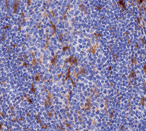 Immunohistochemical analysis of formalin fixed paraffin embedded human tonsil tissue with F5507 at 1:500 dilution.