|
受注:045-509-1970 |
技術サポート:tech@selleck.co.jp 平日9:00〜18:00 1営業日以内にご連絡を差し上げます |
生物学的記述
| Specificity | TCF11/NRF1 Antibody [F22N18] detects endogenous levels of total TCF11/NRF1 protein. |
|---|---|
| Background | TCF11, also known as NRF1 (nuclear factor erythroid 2-related factor 1), is a CNC-bZIP (Cap'n'collar basic leucine zipper) transcription factor belonging to the Nrf family, ubiquitously expressed across tissues and existing as multiple isoforms, including a 120 kDa ER-membrane-bound form and a 65 kDa nuclear-truncated variant. TCF11 features a bZIP domain for DNA binding to antioxidant response elements (AREs), Neh1L (CNC domain with bZIP), Neh2L (ETGE/Neh2-like degron for Keap1 interaction), Neh4L/Neh5L (transactivation domains), and Neh6L (with serine-rich motifs for GSK3 phosphorylation and CRL3-mediated degradation), alongside N-terminal acidic glucose-responsive and ER-targeting domains that enable topological repartitioning. TCF11 translocates from the ER to the nucleus upon proteasome inhibition via p97/VCP-dependent retrotranslocation and proteolytic processing, where it heterodimerizes with small Maf proteins to bind AREs and transcriptionally induce proteasome subunit genes (e.g., PSMB5-8), restoring proteolytic capacity and mitigating proteotoxic stress. This mechanism also regulates mitochondrial biogenesis genes, oxidative stress responses via GCLC, and cytoprotection against rotenone-induced damage, with TCF11 exerting stronger tumor-repressive effects than Nrf1α by upregulating survival genes in hepatocellular carcinoma. Dysregulation links TCF11 to neurodegeneration and cancer progression due to impaired redox/proteostasis homeostasis. |
使用情報
| Application | WB | Dilution |
|
||
|---|---|---|---|---|---|
| Reactivity | Human, Mouse, Monkey | ||||
| Source | Rabbit Monoclonal Antibody | MW | 120-140 kDa | ||
| Storage Buffer | PBS, pH 7.2+50% Glycerol+0.05% BSA+0.01% NaN3 | Storage (from the date of receipt) |
-20°C (avoid freeze-thaw cycles), 2 years | ||
| WB |
Experimental Protocol:
Sample preparation
1. Tissue: Lyse the tissue sample by adding an appropriate volume of ice-cold RIPA/NP-40 Lysis Buffer (containing Protease Inhibitor Cocktail),and homogenize the tissue at a low temperature. 2. Adherent cell: Aspirate the culture medium and wash the cells with ice-cold PBS twice. Lyse the cells by adding an appropriate volume of RIPA/NP-40 Lysis Buffer (containing Protease Inhibitor Cocktail) and put the sample on ice for 5 min. 3. Suspension cell: Transfer the culture medium to a pre-cooled centrifuge tube. Centrifuge and aspirate the supernatant. Wash the cells with ice-cold PBS twice. Lyse the cells by adding an appropriate volume of RIPA/NP-40 Lysis Buffer (containing Protease Inhibitor Cocktail) and put the sample on ice for 5 min. 5. Take a small amount of the lysate to determine the protein concentration; Electrophoretic separation
1. According to the concentration of extracted protein, load appropriate amount of protein sample and marker onto SDS-PAGE gels for electrophoresis. Recommended separating gel (lower gel) concentration: 5%. Reference Table for Selecting SDS-PAGE Separation Gel Concentrations 2. Power up 80V for 30 minutes. Then the power supply is adjusted (110 V~150 V), the Marker is observed, and the electrophoresis can be stopped when the indicator band of the predyed protein Marker where the protein is located is properly separated. (Note that the current should not be too large when electrophoresis, too large current (more than 150 mA) will cause the temperature to rise, affecting the result of running glue. If high currents cannot be avoided, an ice bath can be used to cool the bath.)
Transfer membrane
1. Take out the converter, soak the clip and consumables in the pre-cooled converter;
2. Activate PVDF membrane with methanol for 1 min and rinse with transfer buffer;
3. Install it in the order of "black edge of clip - sponge - filter paper - filter paper - glue -PVDF membrane - filter paper - filter paper - sponge - white edge of clip"; 4. The protein was electrotransferred to PVDF membrane. ( 0.45 µm PVDF membrane is recommended ) Reference Table for Selecting PVDF Membrane Pore Size Specifications Recommended conditions for wet transfer: 200 mA, 120 min. ( Note that the transfer conditions can be adjusted according to the protein size. For high-molecular-weight proteins, a higher current and longer transfer time are recommended. However, ensure that the transfer tank remains at a low temperature to prevent gel melting.)
Block
1. After electrotransfer, wash the film with TBST at room temperature for 5 minutes;
2. Incubate the film in the blocking solution for 1 hour at room temperature;
3. Wash the film with TBST for 3 times, 5 minutes each time.
Antibody incubation
1. Use 5% skim milk powder to prepare the primary antibody working liquid (recommended dilution ratio for primary antibody 1:1000), gently shake and incubate with the film at 4°C overnight; 2. Wash the film with TBST 3 times, 5 minutes each time;
3. Add the secondary antibody to the blocking solution and incubate with the film gently at room temperature for 1 hour;
4. After incubation, wash the film with TBST 3 times for 5 minutes each time.
Antibody staining
1. Add the prepared ECL luminescent substrate (or select other color developing substrate according to the second antibody) and mix evenly;
2. Incubate with the film for 1 minute, remove excess substrate (keep the film moist), wrap with plastic film, and expose in the imaging system.
|
References
|
Application Data
WB
Validated by Selleck
-
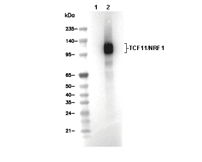 Lane 1: U-2 OS, Lane 2: U-2 OS (MG132, 10 µM, 8 h)