
|
受注:045-509-1970 |
技術サポート:tech@selleck.co.jp 平日9:00〜18:00 1営業日以内にご連絡を差し上げます |
生物学的記述
| Specificity | TET2 Antibody (Rabbit mAb) [H24A10] detects endogenous levels of total TET2 protein. |
|---|---|
| Background | TET2 is a Fe²⁺/α‑ketoglutarate–dependent dioxygenase of the Ten‑Eleven Translocation family that catalyzes stepwise oxidation of 5‑methylcytosine in DNA to 5‑hydroxymethyl‑, 5‑formyl‑ and 5‑carboxylcytosine, thereby coupling DNA methylation dynamics to base‑excision repair and playing a central role in active cytosine demethylation, enhancer function and lineage‑specific gene regulation in hematopoietic and other stem and progenitor cells. The protein has an N‑terminal region that associates with the non‑catalytic CXXC4 domain and a C‑terminal catalytic module composed of a cysteine‑rich segment and a double‑stranded β‑helix fold that coordinates three iron‑binding motifs and one α‑ketoglutarate‑binding site, forming the typical 2‑oxoglutarate oxygenase architecture that positions the methylated cytosine base in the active site for iterative oxidation and enables TET2 to generate 5hmC, 5fC and 5caC at CpG sites within gene bodies and regulatory elements. Genome‑wide and biochemical analyses show that TET2 is highly enriched at enhancers and CpG‑rich promoters involved in hematopoietic differentiation and immune regulation, where its activity shapes the distribution of 5hmC and maintains an open chromatin state permissive for transcription of key regulators of cell cycle, apoptosis, myeloid and lymphoid lineage commitment, and that loss of TET2 function causes promoter and CpG‑island hypermethylation with reduced expression of differentiation‑associated genes. In normal hematopoiesis, TET2 restrains self‑renewal and promotes maturation by modulating enhancer hydroxymethylation in hematopoietic stem and progenitor cells and in germinal center B cells; conditional TET2 deficiency in the B‑cell compartment produces germinal center hyperplasia, impairs plasma cell differentiation and promotes diffuse large B‑cell lymphoma (DLBCL), illustrating that TET2‑dependent enhancer demethylation is required for timely exit from proliferative niches and for humoral immune homeostasis. Somatic loss‑of‑function mutations in TET2 are among the most frequent lesions in myeloid malignancies, including myelodysplastic syndromes, clonal hematopoiesis and AML, and are increasingly recognized in lymphoid neoplasms such as DLBCL, where integrated methylation–expression profiling demonstrates that TET2‑mutant cases display a characteristic hypermethylation signature at CpG islands and CpG‑rich promoters of hematopoietic differentiation genes together with dysregulated transcription networks. Targeted sequencing of DLBCL cohorts has identified TET2 mutations ranging from nonsense and frameshift to missense changes, with frequencies up to ~12% across series and particularly high rates in Epstein–Barr virus–positive DLBCL in elderly patients, where TET2 and LILRB1 are among the most commonly mutated genes, suggesting that TET2 disruption cooperates with viral infection and immune senescence in B‑cell lymphomagenesis. Beyond hematologic disease, reduced TET2 expression and 5hmC levels are reported in several solid tumors such as prostate cancer, melanoma and oral squamous cell carcinoma, and experimental models indicate that TET2 loss contributes to oncogenic epigenetic reprogramming by locking tumor suppressor and differentiation genes in a hypermethylated state and altering enhancer landscapes that control proliferation and invasion. |
使用情報
| Application | WB, ChIP | Dilution |
|
||||
|---|---|---|---|---|---|---|---|
| Reactivity | Mouse | ||||||
| Source | Rabbit Monoclonal Antibody | MW | 224 kDa | ||||
| Storage Buffer | PBS, pH 7.2+50% Glycerol+0.05% BSA+0.01% NaN3 | Storage (from the date of receipt) |
-20°C (avoid freeze-thaw cycles), 2 years | ||||
| WB |
Experimental Protocol:
Sample preparation
1. Tissue: Lyse the tissue sample by adding an appropriate volume of ice-cold RIPA/NP-40 Lysis Buffer (containing Protease Inhibitor Cocktail),and homogenize the tissue at a low temperature or lyse it by sonication on ice, then incubate on ice for 30 minutes. 2. Adherent cell: Aspirate the culture medium and wash the cells with ice-cold PBS twice. Lyse the cells by adding an appropriate volume of RIPA/NP-40 Lysis Buffer (containing Protease Inhibitor Cocktail) , sonicate to lyse the cells, and incubate on ice for 30 minutes. 3. Suspension cell: Transfer the culture medium to a pre-cooled centrifuge tube. Centrifuge and aspirate the supernatant. Wash the cells with ice-cold PBS twice. Lyse the cells by adding an appropriate volume of RIPA/NP-40 Lysis Buffer (containing Protease Inhibitor Cocktail) , sonicate to lyse the cells, and incubate on ice for 30 minutes. 4. Place the lysate into a pre-cooled microcentrifuge tube. Centrifuge at 4°C for 15 min. Collect the supernatant;
5. Remove a small volume of lysate to determine the protein concentration;
6. Combine the lysate with protein loading buffer. Boil 20 µL sample under 95-100°C for 5 min. Centrifuge for 5 min after cool down on ice.
Electrophoretic separation
1. According to the concentration of extracted protein, load appropriate amount of protein sample and marker onto SDS-PAGE gels for electrophoresis. Recommended separating gel (lower gel) concentration: 5%. Reference Table for Selecting SDS-PAGE Separation Gel Concentrations 2. Power up 80V for 30 minutes. Then the power supply is adjusted (110 V~150 V), the Marker is observed, and the electrophoresis can be stopped when the indicator band of the predyed protein Marker where the protein is located is properly separated. (Note that the current should not be too large when electrophoresis, too large current (more than 150 mA) will cause the temperature to rise, affecting the result of running glue. If high currents cannot be avoided, an ice bath can be used to cool the bath.)
Transfer membrane
1. Take out the converter, soak the clip and consumables in the pre-cooled converter;
2. Activate PVDF membrane with methanol for 1 min and rinse with transfer buffer;
3. Install it in the order of "black edge of clip - sponge - filter paper - filter paper - glue -PVDF membrane - filter paper - filter paper - sponge - white edge of clip"; 4. The protein was electrotransferred to PVDF membrane. ( 0.45 µm PVDF membrane is recommended ) Reference Table for Selecting PVDF Membrane Pore Size Specifications Recommended conditions for wet transfer: 250 mA, 180 min. ( Note that the transfer conditions can be adjusted according to the protein size. For high-molecular-weight proteins, a higher current and longer transfer time are recommended. However, ensure that the transfer tank remains at a low temperature to prevent gel melting.)
Block
1. After electrotransfer, wash the film with TBST at room temperature for 5 minutes;
2. Incubate the film in the blocking solution for 1 hour at room temperature;
3. Wash the film with TBST for 3 times, 5 minutes each time.
Antibody incubation
1. Use primary antibody dilution buffer to prepare the primary antibody working liquid (recommended dilution ratio for primary antibody 1:1000), gently shake and incubate with the film at 4°C overnight; 2. Wash the film with TBST 3 times, 5 minutes each time;
3. Add the secondary antibody to the blocking solution and incubate with the film gently at room temperature for 1 hour;
4. After incubation, wash the film with TBST 3 times for 5 minutes each time.
Antibody staining
1. Add the prepared ECL luminescent substrate (or select other color developing substrate according to the second antibody) and mix evenly;
2. Incubate with the film for 1 minute, remove excess substrate (keep the film moist), wrap with plastic film, and expose in the imaging system. (Exposure time of at least 150s is recommended)
|
References
|
Application Data
WB
Validated by Selleck
-
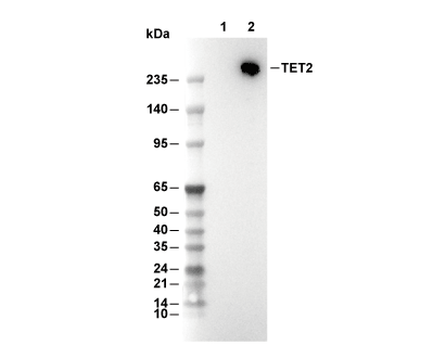 Lane 1: 293T, Lane 2: 293T (mTet2, transfected)