|
受注:045-509-1970 |
技術サポート:tech@selleck.co.jp 平日9:00〜18:00 1営業日以内にご連絡を差し上げます |
生物学的記述
| Specificity | TH1L Antibody [N21A8] detects endogenous levels of total TH1L protein. |
|---|---|
| Background | TH1L (also known as NELF‑C/D) is a metazoan‑specific subunit of the Negative Elongation Factor (NELF) complex, which also includes WHSC2 (NELF‑A), COBRA‑1 (NELF‑B), and NELF‑E and is broadly expressed in mammalian tissues involved in regulated transcription. TH1L, together with COBRA‑1, serves as an integral structural core of the NELF complex that physically links WHSC2 and NELF‑E, enabling assembly of a functional tetrameric NELF complex capable of associating with RNA Polymerase II (RNAPII). The NELF complex cooperates with the DRB‑sensitivity‑inducing factor (DSIF) complex at the promoter‑proximal region of many genes to inhibit RNAPII elongation and establish a paused state, in which the polymerase is halted near the transcription start site and requires additional signaling for productive elongation. Release of RNAPII from this pause is mediated by the positive transcription elongation factor b (p‑TEFb), which phosphorylates NELF (including NELF‑E) and the carboxy‑terminal domain (CTD) of the largest RNAPII subunit, thereby weakening the NELF–RNAPII association and permitting elongation to proceed. Within the NELF complex, TH1L contributes to proper subunit organization and complex stability, and alternative translation initiation at the TH1L locus generates distinct isoforms that may influence complex stoichiometry or localization. TH1L‑containing NELF‑C and NELF‑D isoforms can both support NELF assembly and function, linking TH1L to the regulation of a broad transcriptional program that controls immediate‑early genes, developmental regulators, and stress‑response loci. |
使用情報
| Application | WB, IP, ChIP | Dilution |
|
||||||
|---|---|---|---|---|---|---|---|---|---|
| Reactivity | Human, Mouse, Rat, Monkey | ||||||||
| Source | Rabbit Monoclonal Antibody | MW | 66 kDa | ||||||
| Storage Buffer | PBS, pH 7.2+50% Glycerol+0.05% BSA+0.01% NaN3 | Storage (from the date of receipt) |
-20°C (avoid freeze-thaw cycles), 2 years | ||||||
| WB |
Experimental Protocol:
Sample preparation
1. Tissue: Lyse the tissue sample by adding an appropriate volume of ice-cold RIPA/Nuclear Lysis Buffer (containing Protease Inhibitor Cocktail),and homogenize the tissue at a low temperature. 2. Adherent cell: Aspirate the culture medium and wash the cells with ice-cold PBS twice. Lyse the cells by adding an appropriate volume of RIPA/Nuclear Lysis Buffer (containing Protease Inhibitor Cocktail) and put the sample on ice for 5 min. 3. Suspension cell: Transfer the culture medium to a pre-cooled centrifuge tube. Centrifuge and aspirate the supernatant. Wash the cells with ice-cold PBS twice. Lyse the cells by adding an appropriate volume of RIPA/Nuclear Lysis Buffer (containing Protease Inhibitor Cocktail) and put the sample on ice for 5 min. 4. Place the lysate into a pre-cooled microcentrifuge tube. Centrifuge at 4°C for 15 min. Collect the supernatant;
5. Remove a small volume of lysate to determine the protein concentration;
6. Combine the lysate with protein loading buffer. Boil 20 µL sample under 95-100°C for 5 min. Centrifuge for 5 min after cool down on ice.
Electrophoretic separation
1. According to the concentration of extracted protein, load appropriate amount of protein sample and marker onto SDS-PAGE gels for electrophoresis. Recommended separating gel (lower gel) concentration: 10%. Reference Table for Selecting SDS-PAGE Separation Gel Concentrations 2. Power up 80V for 30 minutes. Then the power supply is adjusted (110 V~150 V), the Marker is observed, and the electrophoresis can be stopped when the indicator band of the predyed protein Marker where the protein is located is properly separated. (Note that the current should not be too large when electrophoresis, too large current (more than 150 mA) will cause the temperature to rise, affecting the result of running glue. If high currents cannot be avoided, an ice bath can be used to cool the bath.)
Transfer membrane
1. Take out the converter, soak the clip and consumables in the pre-cooled converter;
2. Activate PVDF membrane with methanol for 1 min and rinse with transfer buffer;
3. Install it in the order of "black edge of clip - sponge - filter paper - filter paper - glue -PVDF membrane - filter paper - filter paper - sponge - white edge of clip"; 4. The protein was electrotransferred to PVDF membrane. ( 0.45 µm PVDF membrane is recommended ) Reference Table for Selecting PVDF Membrane Pore Size Specifications Recommended conditions for wet transfer: 200 mA, 120 min. ( Note that the transfer conditions can be adjusted according to the protein size. For high-molecular-weight proteins, a higher current and longer transfer time are recommended. However, ensure that the transfer tank remains at a low temperature to prevent gel melting.)
Block
1. After electrotransfer, wash the film with TBST at room temperature for 5 minutes;
2. Incubate the film in the blocking solution for 1 hour at room temperature;
3. Wash the film with TBST for 3 times, 5 minutes each time.
Antibody incubation
1. Use 5% skim milk powder to prepare the primary antibody working liquid (recommended dilution ratio for primary antibody 1:1000), gently shake and incubate with the film at 4°C overnight; 2. Wash the film with TBST 3 times, 5 minutes each time;
3. Add the secondary antibody to the blocking solution and incubate with the film gently at room temperature for 1 hour;
4. After incubation, wash the film with TBST 3 times for 5 minutes each time.
Antibody staining
1. Add the prepared ECL luminescent substrate (or select other color developing substrate according to the second antibody) and mix evenly;
2. Incubate with the film for 1 minute, remove excess substrate (keep the film moist), wrap with plastic film, and expose in the imaging system.
|
References
|
Application Data
WB
Validated by Selleck
-
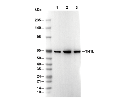 Lane 1: HCT116, Lane 2: RAW264.7, Lane 3: COS-7