|
受注:045-509-1970 |
技術サポート:tech@selleck.co.jp 平日9:00〜18:00 1営業日以内にご連絡を差し上げます |
生物学的記述
| Specificity | Thrombospondin 1 Antibody [N18C17] detects endogenous levels of total Thrombospondin 1 protein. |
|---|---|
| Background | Thrombospondin-1 (TSP-1 or THBS1) is a homotrimeric matricellular glycoprotein secreted by platelets, endothelial cells, and fibroblasts, and integrates into the extracellular matrix to regulate tissue remodeling through a wide array of ligand interactions. TSP-1 consists of an N-terminal globular TSPN domain that forms a β-sandwich of 13 antiparallel strands with a distinctive irregular β4′ segment and a disulfide bond at Cys248, enabling binding to heparin, aggrecan, and integrin αvβ3. This is followed by a von Willebrand factor C domain, three type I repeats (TSRs) characterized by stacked tryptophan/arginine ladders and conserved WXXWCSXG motifs that mediate engagement with CD36 and CD47 and inhibit MMP2/9, EGF-like type II repeats, and 15 calponin homology repeats with an RGD motif in the last repeat conferring affinity for integrins αvβ3 and αIIbβ3. The protein terminates with a C-terminal lectin-like globular domain that binds proteoglycans and stabilizes oligomers through interchain disulfide bonds at Cys252 and Cys256. TSP-1 potently inhibits angiogenesis by clustering CD47, thereby disrupting VEGF-R2 and nitric oxide (NO) signaling via SHP-2 phosphatase recruitment and blockade of eNOS S-nitrosylation. TSP-1 also activates latent TGF-β1 through integrin αvβ3/β8-mediated, shear-dependent unfolding of the latency-associated peptide, leading to Smad2/3 phosphorylation and fibrosis. It induces anoikis and CD36-mediated apoptosis in endothelial and smooth muscle cells via caspase-8/3 cascades and inhibition of Fyn/FAK, while also paradoxically promoting leukocyte transmigration and phagocytosis through calreticulin/LRP1/CD91-mediated engulfment of apoptotic bodies or pathogens. TSP-1 deficiency exacerbates tumorigenesis by permitting pathological angiogenesis and immune evasion in models of breast and prostate cancer, while its 3TSR fragment possesses strong anti-angiogenic activity when released by ADAMTS1 cleavage. |
使用情報
| Application | WB, IP | Dilution |
|
||||
|---|---|---|---|---|---|---|---|
| Reactivity | Human, Mouse, Rat | ||||||
| Source | Rabbit Monoclonal Antibody | MW | 170 kDa | ||||
| Storage Buffer | PBS, pH 7.2+50% Glycerol+0.05% BSA+0.01% NaN3 | Storage (from the date of receipt) |
-20°C (avoid freeze-thaw cycles), 2 years | ||||
| WB |
Experimental Protocol:
Sample preparation
1. Tissue: Lyse the tissue sample by adding an appropriate volume of ice-cold RIPA/NP-40 Lysis Buffer (containing Protease Inhibitor Cocktail),and homogenize the tissue at a low temperature. 2. Adherent cell: Aspirate the culture medium and wash the cells with ice-cold PBS twice. Lyse the cells by adding an appropriate volume of RIPA/NP-40 Lysis Buffer (containing Protease Inhibitor Cocktail) and put the sample on ice for 5 min. 3. Suspension cell: Transfer the culture medium to a pre-cooled centrifuge tube. Centrifuge and aspirate the supernatant. Wash the cells with ice-cold PBS twice. Lyse the cells by adding an appropriate volume of RIPA/NP-40 Lysis Buffer (containing Protease Inhibitor Cocktail) and put the sample on ice for 5 min. 4. Place the lysate into a pre-cooled microcentrifuge tube. Centrifuge at 4°C for 15 min. Collect the supernatant;
5. Remove a small volume of lysate to determine the protein concentration;
6. Combine the lysate with protein loading buffer. Boil 20 µL sample under 95-100°C for 5 min. Centrifuge for 5 min after cool down on ice.
Electrophoretic separation
1. According to the concentration of extracted protein, load appropriate amount of protein sample and marker onto SDS-PAGE gels for electrophoresis. Recommended separating gel (lower gel) concentration: 5%. Reference Table for Selecting SDS-PAGE Separation Gel Concentrations 2. Power up 80V for 30 minutes. Then the power supply is adjusted (110 V~150 V), the Marker is observed, and the electrophoresis can be stopped when the indicator band of the predyed protein Marker where the protein is located is properly separated. (Note that the current should not be too large when electrophoresis, too large current (more than 150 mA) will cause the temperature to rise, affecting the result of running glue. If high currents cannot be avoided, an ice bath can be used to cool the bath.)
Transfer membrane
1. Take out the converter, soak the clip and consumables in the pre-cooled converter;
2. Activate PVDF membrane with methanol for 1 min and rinse with transfer buffer;
3. Install it in the order of "black edge of clip - sponge - filter paper - filter paper - glue -PVDF membrane - filter paper - filter paper - sponge - white edge of clip"; 4. The protein was electrotransferred to PVDF membrane. ( 0.45 µm PVDF membrane is recommended ) Reference Table for Selecting PVDF Membrane Pore Size Specifications Recommended conditions for wet transfer: 200 mA, 120 min. ( Note that the transfer conditions can be adjusted according to the protein size. For high-molecular-weight proteins, a higher current and longer transfer time are recommended. However, ensure that the transfer tank remains at a low temperature to prevent gel melting.)
Block
1. After electrotransfer, wash the film with TBST at room temperature for 5 minutes;
2. Incubate the film in the blocking solution for 1 hour at room temperature;
3. Wash the film with TBST for 3 times, 5 minutes each time.
Antibody incubation
1. Use 5% skim milk powder to prepare the primary antibody working liquid (recommended dilution ratio for primary antibody 1:1000), gently shake and incubate with the film at 4°C overnight; 2. Wash the film with TBST 3 times, 5 minutes each time;
3. Add the secondary antibody to the blocking solution and incubate with the film gently at room temperature for 1 hour;
4. After incubation, wash the film with TBST 3 times for 5 minutes each time.
Antibody staining
1. Add the prepared ECL luminescent substrate (or select other color developing substrate according to the second antibody) and mix evenly;
2. Incubate with the film for 1 minute, remove excess substrate (keep the film moist), wrap with plastic film, and expose in the imaging system.
|
References
|
Application Data
WB
Validated by Selleck
-
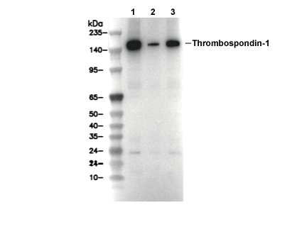 Lane 1: ACHN, Lane 2: LN18, Lane 3: MEF