
|
受注:045-509-1970 |
技術サポート:tech@selleck.co.jp 平日9:00〜18:00 1営業日以内にご連絡を差し上げます |
生物学的記述
| Specificity | TWIST1 Antibody (Rabbit mAb) [M21K22] detects endogenous levels of total TWIST1 protein. |
|---|---|
| Background | TWIST1 belongs to the class B basic helix-loop-helix family of transcription factors and functions as a master regulator of mesoderm formation and mesenchymal differentiation during embryonic morphogenesis, acting through its bHLH domain to bind E-box regulatory sequences on target gene promoters. The protein forms both homodimers and heterodimers, with the heterodimer between TWIST1 and the class A bHLH factor E12 representing the predominant transcriptionally active complex driving prometastatic gene programs, while homodimerization with itself or with other class B partners such as HAND2 produces functionally distinct outcomes; dimer choice within the cell is governed largely by the relative availability of these bHLH partners and by inhibitory Id proteins, which sequester E12 through competitive binding and thereby actively reshape the pool of TWIST1 complexes able to engage DNA. Heterodimerization with E12 depends on coordinated phosphorylation, since a phosphorylation-deficient TWIST1 mutant at serine 68 selectively impairs TWIST1-E12 heterodimer formation and downstream target gene induction while leaving TWIST1 homodimer formation intact, demonstrating that phosphorylation state functions as a molecular switch controlling which dimer species predominates and, consequently, which transcriptional program is executed. Once assembled, the TWIST1-E12 heterodimer drives epithelial-mesenchymal transition by directly repressing E-cadherin expression, a repression achieved through recruitment of the Mi-2/NuRD nucleosome remodeling and deacetylase complex via interactions with MTA2 and RbAp46, coupling TWIST1 DNA binding to active chromatin remodeling rather than passive transcriptional interference. TWIST1 additionally engages the TWIST-box domain, a separate conserved region that binds the runt domain of RUNX2 and inhibits its transactivation activity, while TWIST1 also directly represses RUNX2 expression, together restraining premature osteoblast differentiation and positioning TWIST1 as a checkpoint over osteogenic commitment during skeletal development. In cancer contexts, TWIST1-driven dissemination of epithelial cells can proceed while cytokeratin expression and epithelial identity are preserved, with TWIST1 instead reprogramming extracellular matrix and cell-matrix adhesion genes rather than cell-cell adhesion genes, indicating that TWIST1 promotes tumor cell dissemination through a mechanism broader than E-cadherin loss alone. TWIST1 expression is elevated across breast, liver, prostate, and gastric cancers and is upregulated by multiple upstream inputs including STAT3, HIF-1α, and NF-κB, correlating with invasive and metastatic phenotypes, while heterozygous loss-of-function mutations affecting the bHLH domain cause Saethre-Chotzen syndrome, a craniosynostosis disorder, establishing TWIST1 dosage and dimerization fidelity as critical parameters across both developmental and oncogenic contexts. |
使用情報
| Application | IHC | Dilution |
|
||
|---|---|---|---|---|---|
| Reactivity | Mouse | ||||
| Source | Rabbit Monoclonal Antibody | MW | 21 kDa | ||
| Storage Buffer | PBS, pH 7.2+50% Glycerol+0.05% BSA+0.01% NaN3 | Storage (from the date of receipt) |
-20°C (avoid freeze-thaw cycles), 2 years | ||
| IHC |
Experimental Protocol:
Deparaffinization/Rehydration
1. Deparaffinize/hydrate sections:
2. Incubate sections in three washes of xylene for 5 min each.
3. Incubate sections in two washes of 100% ethanol for 10 min each.
4. Incubate sections in two washes of 95% ethanol for 10 min each.
5. Wash sections two times in dH2O for 5 min each.
6.Antigen retrieval: For Citrate: Heat slides in a microwave submersed in 1X citrate unmasking solution until boiling is initiated; continue with 10 min at a sub-boiling temperature (95°-98°C). Cool slides on bench top for 30 min.
Staining
1. Wash sections in dH2O three times for 5 min each.
2. Incubate sections in 3% hydrogen peroxide for 10 min.
3. Wash sections in dH2O two times for 5 min each.
4. Wash sections in wash buffer for 5 min.
5. Block each section with 100–400 µl of blocking solution for 1 hr at room temperature.
6. Remove blocking solution and add 100–400 µl primary antibody diluent in to each section. Incubate overnight at 4°C.
7. Remove antibody solution and wash sections with wash buffer three times for 5 min each.
8. Cover section with 1–3 drops HRPas needed. Incubate in a humidified chamber for 30 min at room temperature.
9. Wash sections three times with wash buffer for 5 min each.
10. Add DAB Chromogen Concentrate to DAB Diluent and mix well before use.
11. Apply 100–400 µl DAB to each section and monitor closely. 1–10 min generally provides an acceptable staining intensity.
12. Immerse slides in dH2O.
13. If desired, counterstain sections with hematoxylin.
14. Wash sections in dH2O two times for 5 min each.
15. Dehydrate sections: Incubate sections in 95% ethanol two times for 10 sec each; Repeat in 100% ethanol, incubating sections two times for 10 sec each; Repeat in xylene, incubating sections two times for 10 sec each.
16. Mount sections with coverslips and mounting medium.
|
References
|
Application Data
IHC
Validated by Selleck
-
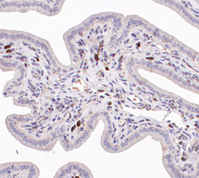 Immunohistochemical analysis of formalin fixed paraffin embedded mouse uterine tissue with F5951 at 1:250 dilution.