|
受注:045-509-1970 |
技術サポート:tech@selleck.co.jp 平日9:00〜18:00 1営業日以内にご連絡を差し上げます |
生物学的記述
| Specificity | VEGF165b Antibody [B10E21] detects endogenous levels of total VEGF165b protein. |
|---|---|
| Background | VEGF165b arises from alternative splicing of exon 8 in the vascular endothelial growth factor A (VEGF-A) gene, differing from the proangiogenic VEGF165a isoform by six amino acids at the C-terminus—VEGF165a terminating with the sequence CDKPRR, while VEGF165b possesses the terminal sequence SLTRKD derived from the distal splice acceptor site in exon 8b. This structural divergence alters receptor binding properties and downstream signaling consequences without changing the overall topology of receptor engagement, positioning VEGF165b as an endogenous regulator that modulates rather than completely blocks VEGF-mediated angiogenesis. VEGF165b functions as an antagonist of proangiogenic VEGF165a by preferentially binding to VEGF receptor 1 (VEGFR1) over VEGFR2, preventing VEGF165a from activating the VEGFR1-STAT3 signaling axis that drives angiogenesis and perfusion recovery in peripheral arterial disease (PAD) and other ischemic conditions. The protein exhibits elevated expression in human PAD muscle biopsies compared to control tissues, with higher binding affinity to VEGFR1 relative to VEGF165a, and VEGF165b levels correlate inversely with VEGFR1 activation status but not VEGFR2 activation, challenging the traditional view that VEGF165b primarily antagonizes VEGFR2 signaling. Inhibition of VEGF165b through isoform-specific monoclonal antibodies enhances perfusion recovery in severe PAD without activating VEGFR2 signaling, instead increasing VEGFR1 activation and promoting VEGFR1-STAT3 binding interactions that trigger STAT3 phosphorylation and nuclear translocation independent of Janus-activated kinase 1/2 (JAK1/JAK2) activation. The protein binds VEGFR2 with similar affinity to VEGF165a but fails to induce receptor phosphorylation and downstream mitogen-activated protein kinase (MAPK) and phospholipase C gamma (PLCγ) activation, instead competitively inhibiting VEGF165a-mediated VEGFR2 autophosphorylation at tyrosine residues Y1175 and Y1214, which are essential for endothelial cell proliferation and migration. VEGF165b inhibits VEGF165a-stimulated endothelial cell proliferation, migration, and capillary tube formation with an IC50 approximately tenfold lower than the concentration of VEGF165a present, demonstrating potent inhibitory capacity even when expressed at substoichiometric levels relative to proangiogenic isoforms. The splice variant exhibits downregulation in multiple human malignancies, including renal cell carcinoma, prostate cancer, and melanoma, with loss of VEGF165b expression correlating with increased tumor angiogenesis, progression, and metastatic potential, while exogenous VEGF165b administration inhibits tumor growth and vascular density. VEGF165b expression becomes regulated at the level of alternative splicing through factors including transforming growth factor-beta 1 (TGF-β1) and insulin-like growth factor 1 (IGF-1), which promote proximal splice site selection favoring VEGF165a production, whereas serine-arginine-rich splicing factor 1 (SRSF1) promotes distal splice site usage, generating VEGF165b, positioning splicing factor availability and activity as determinants of the VEGF165a/VEGF165b expression ratio. The protein regulates immune cell function beyond endothelial effects, augmenting natural killer cell cytolytic activity against leukemia cells through upregulation of perforin and granzyme B expression via VEGFR1-phospholipase C (PLC) pathway activation, demonstrating immunomodulatory capabilities distinct from simple anti-angiogenic mechanisms. VEGF165b exhibits tissue-specific expression patterns with high levels detected in healthy kidney glomeruli and retinal pigment epithelium, maintaining basal vascular quiescence and preventing pathological neovascularization, while reduced expression in diabetic retinopathy and age-related macular degeneration associates with aberrant angiogenesis. |
使用情報
| Application | WB, IHC, IF | Dilution |
|
||||||||||||
|---|---|---|---|---|---|---|---|---|---|---|---|---|---|---|---|
| Reactivity | Human | ||||||||||||||
| Source | Mouse Monoclonal Antibody | MW | 44 kDa | ||||||||||||
| Storage Buffer | PBS, pH 7.2+50% Glycerol+0.05% BSA+0.01% NaN3 | Storage (from the date of receipt) |
-20°C (avoid freeze-thaw cycles), 2 years | ||||||||||||
References
|
Application Data
IHC
Validated by Selleck
-
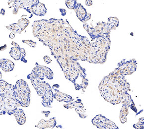 Immunohistochemical analysis of formalin fixed paraffin embedded human placenta tissue with F5992 at 1:50 dilution.