
|
受注:045-509-1970 |
技術サポート:tech@selleck.co.jp 平日9:00〜18:00 1営業日以内にご連絡を差し上げます |
生物学的記述
| Specificity | VPS15 Antibody (Rabbit mAb) [A22G22] detects endogenous levels of total VPS15 protein. |
|---|---|
| Background | VPS15 (also known as p150) is a large HEAT‑repeat scaffold and regulatory subunit of the class III phosphatidylinositol 3‑kinase complexes PI3KC3‑C1 and PI3KC3‑C2, where it associates with VPS34 and Beclin 1 and additional subunits such as ATG14L or UVRAG/BIF‑1 to control localized synthesis of phosphatidylinositol 3‑phosphate and thereby direct membrane trafficking events in autophagy, endocytosis and cytokinesis. The N‑terminal region of VPS15 contains a putative protein kinase–like domain that is essential for VPS34 activation, followed by HEAT repeats and a C‑terminal seven‑bladed WD40 β‑propeller that bridges VPS15–VPS34 and VPS30/Beclin‑1–UVRAG or VPS38 pairs in the endosomal PI3KC3‑C2 complex, creating an extended architecture which positions the catalytic VPS34 lipid kinase close to specific endosomal, autophagosomal or cytokinetic membranes and couples enzyme activity to cargo selection and complex assembly. In the autophagic PI3KC3‑C1 complex, VPS15 cooperates with VPS34, Beclin 1 and ATG14L to generate PI3P at phagophore initiation sites, recruiting downstream effectors such as WIPI proteins and DFCP1 that mark omegasomes and support LC3 conjugation and autophagosome formation; in PI3KC3‑C2, VPS15 forms a sub‑complex with VPS34, Beclin 1, UVRAG and BIF‑1 that regulates degradative endocytic trafficking and cytokinesis, with high‑content microscopy showing that depletion of VPS15 or UVRAG/BIF‑1 impairs growth factor receptor degradation and completion of cell division and that UVRAG and BIF‑1 localize strongly to the midbody during cytokinesis. VPS15 is indispensable for VPS34 activity, and conserved complexes containing VPS34 and VPS15 promote early autophagosome formation in Drosophila and mammalian cells, with loss of VPS15 reducing PI3P generation, blocking autophagosome biogenesis and preventing autophagic clearance of protein aggregates, emphasizing VPS15’s central role in the autophagic PI3P axis. Phosphoproteomic studies identify VPS15 as a direct substrate of the ULK kinase complex that initiates autophagy: ULK‑mediated phosphorylation of VPS15 modulates PI3KC3‑C1 activity and autophagic flux, placing VPS15 at the intersection between upstream nutrient‑sensing ULK signaling and downstream VPS34‑dependent membrane remodeling. Recent cryo‑EM work on PI3KC3 regulation further reveals a structural pathway by which VPS15’s HEAT‑repeat arm and WD40 domain transmit conformational changes to VPS34 and Beclin 1 upon binding regulatory inputs, enabling allosteric control of lipid kinase activity in response to stress, growth factor withdrawal or endocytic cues. In mammalian cells, a PI3KC3‑C2 sub‑complex containing VPS15 and VPS34 is recruited by ubiquitinated transferrin receptor to LC3‑positive recycling endosome membranes, where VPS15 supports PI3P synthesis that coordinates both LC3 lipidation and subsequent ESCRT‑mediated phagophore closure, linking VPS15 activity directly to the sequential steps of autophagosome formation and maturation. VPS15 deficiency or mutation has been associated with defects in stress‑induced and developmental autophagy, impaired receptor downregulation, abnormal cytokinesis and neurodegenerative phenotypes in Mendelian disease studies, consistent with a broad requirement for VPS15‑regulated PI3KC3 complexes in endosomal sorting, lysosomal degradation and neuronal homeostasis. |
使用情報
| Application | WB, IHC | Dilution |
|
||||
|---|---|---|---|---|---|---|---|
| Reactivity | Mouse, Rat, Human | ||||||
| Source | Rabbit Monoclonal Antibody | MW | 153 kDa | ||||
| Storage Buffer | PBS, pH 7.2+50% Glycerol+0.05% BSA+0.01% NaN3 | Storage (from the date of receipt) |
-20°C (avoid freeze-thaw cycles), 2 years | ||||
| WB |
Experimental Protocol:
Sample preparation
1. Tissue: Lyse the tissue sample by adding an appropriate volume of ice-cold RIPA/NP-40 Lysis Buffer (containing Protease Inhibitor Cocktail),and homogenize the tissue at a low temperature or lyse it by sonication on ice, then incubate on ice for 30 minutes. 2. Adherent cell: Aspirate the culture medium and wash the cells with ice-cold PBS twice. Lyse the cells by adding an appropriate volume of RIPA/NP-40 Lysis Buffer (containing Protease Inhibitor Cocktail) , sonicate to lyse the cells, and incubate on ice for 30 minutes. 3. Suspension cell: Transfer the culture medium to a pre-cooled centrifuge tube. Centrifuge and aspirate the supernatant. Wash the cells with ice-cold PBS twice. Lyse the cells by adding an appropriate volume of RIPA/NP-40 Lysis Buffer (containing Protease Inhibitor Cocktail) , sonicate to lyse the cells, and incubate on ice for 30 minutes. 4. Place the lysate into a pre-cooled microcentrifuge tube. Centrifuge at 4°C for 15 min. Collect the supernatant;
5. Remove a small volume of lysate to determine the protein concentration;
6. Combine the lysate with protein loading buffer. Boil 20 µL sample under 95-100°C for 5 min. Centrifuge for 5 min after cool down on ice.
Electrophoretic separation
1. According to the concentration of extracted protein, load appropriate amount of protein sample and marker onto SDS-PAGE gels for electrophoresis. Recommended separating gel (lower gel) concentration: 5%. Reference Table for Selecting SDS-PAGE Separation Gel Concentrations 2. Power up 80V for 30 minutes. Then the power supply is adjusted (110 V~150 V), the Marker is observed, and the electrophoresis can be stopped when the indicator band of the predyed protein Marker where the protein is located is properly separated. (Note that the current should not be too large when electrophoresis, too large current (more than 150 mA) will cause the temperature to rise, affecting the result of running glue. If high currents cannot be avoided, an ice bath can be used to cool the bath.)
Transfer membrane
1. Take out the converter, soak the clip and consumables in the pre-cooled converter;
2. Activate PVDF membrane with methanol for 1 min and rinse with transfer buffer;
3. Install it in the order of "black edge of clip - sponge - filter paper - filter paper - glue -PVDF membrane - filter paper - filter paper - sponge - white edge of clip"; 4. The protein was electrotransferred to PVDF membrane. ( 0.45 µm PVDF membrane is recommended ) Reference Table for Selecting PVDF Membrane Pore Size Specifications Recommended conditions for wet transfer: 200 mA, 120 min. ( Note that the transfer conditions can be adjusted according to the protein size. For high-molecular-weight proteins, a higher current and longer transfer time are recommended. However, ensure that the transfer tank remains at a low temperature to prevent gel melting.)
Block
1. After electrotransfer, wash the film with TBST at room temperature for 5 minutes;
2. Incubate the film in the blocking solution for 1 hour at room temperature;
3. Wash the film with TBST for 3 times, 5 minutes each time.
Antibody incubation
1. Use primary antibody dilution buffer to prepare the primary antibody working liquid (recommended dilution ratio for primary antibody 1:1000), gently shake and incubate with the film at 4°C overnight; 2. Wash the film with TBST 3 times, 5 minutes each time;
3. Add the secondary antibody to the blocking solution and incubate with the film gently at room temperature for 1 hour;
4. After incubation, wash the film with TBST 3 times for 5 minutes each time.
Antibody staining
1. Add the prepared ECL luminescent substrate (or select other color developing substrate according to the second antibody) and mix evenly;
2. Incubate with the film for 1 minute, remove excess substrate (keep the film moist), wrap with plastic film, and expose in the imaging system.
|
| IHC |
Experimental Protocol:
Deparaffinization/Rehydration
1. Deparaffinize/hydrate sections:
2. Incubate sections in three washes of xylene for 5 min each.
3. Incubate sections in two washes of 100% ethanol for 10 min each.
4. Incubate sections in two washes of 95% ethanol for 10 min each.
5. Wash sections two times in dH2O for 5 min each.
6.Antigen retrieval: For Citrate: Heat slides in a microwave submersed in 1X citrate unmasking solution until boiling is initiated; continue with 10 min at a sub-boiling temperature (95°-98°C). Cool slides on bench top for 30 min.
Staining
1. Wash sections in dH2O three times for 5 min each.
2. Incubate sections in 3% hydrogen peroxide for 10 min.
3. Wash sections in dH2O two times for 5 min each.
4. Wash sections in wash buffer for 5 min.
5. Block each section with 100–400 µl of blocking solution for 1 hr at room temperature.
6. Remove blocking solution and add 100–400 µl primary antibody diluent in to each section. Incubate overnight at 4°C.
7. Remove antibody solution and wash sections with wash buffer three times for 5 min each.
8. Cover section with 1–3 drops HRPas needed. Incubate in a humidified chamber for 30 min at room temperature.
9. Wash sections three times with wash buffer for 5 min each.
10. Add DAB Chromogen Concentrate to DAB Diluent and mix well before use.
11. Apply 100–400 µl DAB to each section and monitor closely. 1–10 min generally provides an acceptable staining intensity.
12. Immerse slides in dH2O.
13. If desired, counterstain sections with hematoxylin.
14. Wash sections in dH2O two times for 5 min each.
15. Dehydrate sections: Incubate sections in 95% ethanol two times for 10 sec each; Repeat in 100% ethanol, incubating sections two times for 10 sec each; Repeat in xylene, incubating sections two times for 10 sec each.
16. Mount sections with coverslips and mounting medium.
|
References
|
Application Data
WB
Validated by Selleck
-
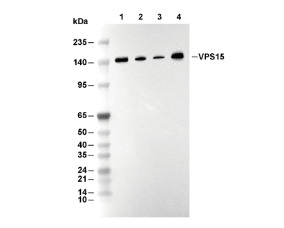 Lane 1: Raji, Lane 2: Jurkat, Lane 3: MOLT4, Lane 4: C6