
- 阻害剤
- 研究分野別
- PI3K/Akt/mTOR
- Epigenetics
- Methylation
- Immunology & Inflammation
- Protein Tyrosine Kinase
- Angiogenesis
- Apoptosis
- Autophagy
- ER stress & UPR
- JAK/STAT
- MAPK
- Cytoskeletal Signaling
- Cell Cycle
- TGF-beta/Smad
- 化合物ライブラリー
- 抗体
- 新製品
- お問い合わせ
E7080 (Lenvatinib)
別名:E7080
レンバチニブ (Lenvatinib (E7080)) はマルチターゲット阻害剤の一種であり、主に VEGFR2(KDR)/VEGFR3(Flt-4) に対して活性を示し IC50 は 4 nM/5.2 nMである一方、VEGFR1/Flt-1に対しては活性が弱く、cell-free assay において FGFR1, PDGFRα/βよりも VEGFR2/3 に対して 10 倍高い選択性を示します。レンバチニブ (E7080) はまた FGFR1-4, PDGFR, Kit (c-Kit), RET (c-RET) も阻害し、強力な抗腫瘍活性を呈します。
CAS No. 417716-92-8
文献中Selleckの製品使用例(141)
製品安全説明書
現在のバッチを見る:
純度:
99.97%
99.97
E7080 (Lenvatinib)と併用されることが多い化合物
Padnarsertib (KPT-9274, ATG-019)
It and KPT 9274 combination exhibit superior anti-tumor activity in anaplastic thyroid cancer cells (8505C) subcutaneous xenografts.
E7080 (Lenvatinib)関連製品
シグナル伝達経路
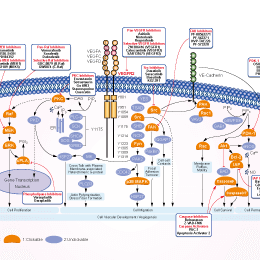
VEGFR阻害剤の選択性比較
Cell Data
| Cell Lines | Assay Type | Concentration | Incubation Time | 活性情報 | PMID |
|---|---|---|---|---|---|
| HT29 cells | Cytotoxicity assay | 25, 50 nM | 72 h | cytotoxic dose: 50 nM and noncytotoxic dose: 25 nM | 24815456 |
| ATC cells | Function assay | 1, 25 and 50 μM | 72 h | Phosphorylated/non-phosphorylated Akt or ERK1/2 proteins (evaluated by ELISA) in lenvatinib-treated samples were significantly reduced in ATC cell cultures. | 29517103 |
| TPC-1 and K1 cells | Function assay | 50 μM | 24 h | The inhibitory effects of lenvatinib on the viability of both cell lines were not influenced by the leptin treatment. | 30906321 |
| DX3 and U2OS cells | Function assay | 1 μM and 10 μM | 16 hours | Lenvatinib inhibit tumor cells migration and invasion at concentrations that both inhibit its known targets and are achievable clinically. | 21781317 |
| HCC cell lines Hep3B2.1-7, HuH-7, and JHH-7 | Proliferation assay | 6 days | Lenvatinib showed selective and potent antiproliferative activity against the HCC cell lines Hep3B2.1‐7, HuH‐7, and JHH‐7, with IC50 values of 0.23, 0.42, and 0.64 μmol/L, respectively. | 29733511 | |
| 他の多くの細胞株試験データをご覧になる場合はこちらをクリックして下さい | |||||
生物活性
| 製品説明 | レンバチニブ (Lenvatinib (E7080)) はマルチターゲット阻害剤の一種であり、主に VEGFR2(KDR)/VEGFR3(Flt-4) に対して活性を示し IC50 は 4 nM/5.2 nMである一方、VEGFR1/Flt-1に対しては活性が弱く、cell-free assay において FGFR1, PDGFRα/βよりも VEGFR2/3 に対して 10 倍高い選択性を示します。レンバチニブ (E7080) はまた FGFR1-4, PDGFR, Kit (c-Kit), RET (c-RET) も阻害し、強力な抗腫瘍活性を呈します。 | |||||||||||
|---|---|---|---|---|---|---|---|---|---|---|---|---|
| Targets |
|
| In Vitro | ||||
| In vitro |
E7080, as a potent inhibitor of in vitro angiogenesis, shows a significantly inhibitory effect on VEGF/KDR and SCF/Kit signaling. According to the in vitro receptor tyrosine and serine/threonine kinase assays, this compound inhibits Flt-1, KDR, Flt-4 with IC50 of 22, 4.0 and 5.2 nM, respectively. In addition to these kinases, this compound also inhibits FGFR1 and PDGFR tyrosine kinases with IC50 value of 46, 51 and 100 nM for FGFR1, PDGFRα and PDGFRβ, respectively. This compound potently inhibits phosphorylation of VEGFR2 (IC50, 0.83 nM) and VEGFR3 (IC50, 0.36 nM) in HUVECs which is stimulated by VEGF and VEGF-C, respectively. A recent study shows that this compound treatment (both at 1 μM and 10 μM) results in a significant inhibition of cell migration and invasion by inhibiting FGFR and PDGFR signaling. |
|||
|---|---|---|---|---|
| Kinase Assay | In vitro kinase assay | |||
| Tyrosine kinase assays are performed by HTRF (KDR, VEGFR1, FGFR1, c-Met, EGFR) and ELISA (PDGFRβ), using the recombinant kinase domains of receptors. In both assays, 4 μL of serial dilutions of this compound are mixed in a 96-well round plate with 10 μL of enzyme, 16 μL of poly (GT) solution (250 ng) and 10 μL of ATP solution (1 μM ATP) (final concentration of DMSO is 0.1%). In wells for blanks, no enzyme is added. In control wells no test article is added. The kinase reaction is initiated by adding ATP solution to each well. After 30-minute incubation at 30°C, the reaction is stopped by adding 0.5 M EDTA (10 μL/well) to the reaction mixture in each well. Dilution buffer adequate to each kinase assay is added to the reaction mixture. In the HTRF assay, 50 μL of the reaction mixture is transferred to a 96-well 1/2 area black EIA/RIA plate, HTRF solution (50 μL/well) is added to the reaction mixture, and then kinase activity is determined by measurement of fluorescence with a time-resolved fluorescence detector at an excitation wavelength of 337 nm and an emission wavelengths of 620 and 665 nm. In the ELISA, 50 μL of the reaction mixture is incubated in avidin coated 96-well polystyrene plates at room temperature for 30 minutes. After washing with wash buffer, PY20-HRP solution (70 μL/well) is added and the reaction mixture is incubated at room temperature for 30 minutes. After washing with wash buffer, TMB reagent (100 μL/well) is added to each well. After several minutes (10–30 minutes), 1 M H3PO4 (100 μL/well) is added to each well. Kinase activity is determined by measurement of absorbance at 450 nm with a microplate reader. | ||||
| 細胞実験 | 細胞株 | HUVECs | ||
| 濃度 | 0-10 μM | |||
| 反応時間 | 72 hours | |||
| 実験の流れ | HUVECs (1,000 cells in each well in serum-free medium containing 2% fetal bovine serum) and L6 rat skeletal muscle myoblasts (5,000 cells in each well in serum-free DMEM) are dispensed in a 96-well plate and incubated overnight. This compound and either VEGF (20 ng/mL) or FGF-2 (20 ng/mL) containing 2% fetal bovine serum and PDGFβ (40 ng/mL) are added to each well. Cells are incubated for 3 days and then the ratios of surviving cells are measured by WST-1 reagent. For proliferation assay, samples are duplicated and three separate experiments are done. |
|||
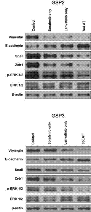
| 実験結果図 | Methods | Biomarkers | 結果図 | PMID |
| Western blot | Vimentin / E-cadherin / Snail / Zeb1 β-catenin Ki-67 / Cyclin D1 / CDK4 / p21 / p53 / Apaf-1 / p-NFκB / Bcl-2 / Cleaved-caspase 3 phospho-RET phospho-FGFR1 / FGFR1 /phospho-FRS2 / FRS2 / phospho-MEK / phospho-ERK |
|
30286728 | |
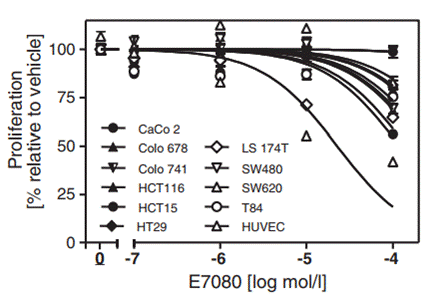
| Growth inhibition assay | Cell viability |
|
25425971 | |
| In Vivo | ||
| In Vivo |
When orally administrated in a H146 xenograft model, Lenvatinib (E7080) inhibits the growth of H146 tumor at 30 and 100 mg/kg in a dose-dependent manner and leads to tumor regression at 100 mg/kg. Furthermore, this compound at 100 mg/kg decreases microvessel density more than anti-VEGF antibody and imatinib treatment. It significantly inhibits local tumor growth in a MDA-MB-231 mammary fat pad (m.f.p.) model with RTVs (calculated tumor volume on day 8/tumor volume on day 1) of 0.81, and reduces both angiogenesis and lymphangiogenesis of established metastatic nodules of MDA-MB-231 tumor in the lymph nodes. |
|
|---|---|---|
| 動物実験 | 動物モデル | H146 tumor cells are implanted subcutaneously (s.c.) into the flank region of female BALB/c nude mice. |
| 投与量 | ≤100 mg/kg | |
| 投与経路 | Administered via p.o. | |
| NCT Number | Recruitment | Conditions | Sponsor/Collaborators | Start Date | Phases |
|---|---|---|---|---|---|
| NCT06161558 | Not yet recruiting | Neoplasms |
National Cancer Institute (NCI)|National Institutes of Health Clinical Center (CC) |
May 15 2024 | Phase 1 |
| NCT05846724 | Not yet recruiting | Kaposi Sarcoma|Classic Kaposi Sarcoma|Refractory Kaposi Sarcoma |
Fondazione IRCCS Ca'' Granda Ospedale Maggiore Policlinico |
February 1 2024 | Phase 2 |
| NCT05903833 | Not yet recruiting | Recurrent Vulvar Cancer|Persistent Vulvar Cancer|Metastatic Vulva Cancer|Locally Advanced Vulvar Cancer |
AGO Research GmbH |
January 1 2024 | Phase 2 |
| NCT05901194 | Not yet recruiting | Hepatocellular Carcinoma Non-resectable |
Assistance Publique - Hôpitaux de Paris|Laboratoire EISAI |
June 2023 | Phase 1|Phase 2 |
|
化学情報
| 分子量 | 426.85 | 化学式 | C21H19ClN4O4 |
| CAS No. | 417716-92-8 | SDF | Download E7080 (Lenvatinib) SDFをダウンロードする |
| Smiles | COC1=CC2=NC=CC(=C2C=C1C(=O)N)OC3=CC(=C(C=C3)NC(=O)NC4CC4)Cl | ||
| 保管 | |||
|
In vitro |
DMSO : 20 mg/mL ( (46.85 mM); 吸湿したDMSOは溶解度を減少させます。新しいDMSOをご使用ください。) Water : Insoluble Ethanol : Insoluble |
モル濃度計算器 |
|
in vivo Add solvents to the product individually and in order. |
投与溶液組成計算機 | |||||
実験計算
投与溶液組成計算機(クリア溶液)
ステップ1:実験データを入力してください。(実験操作によるロスを考慮し、動物数を1匹分多くして計算・調製することを推奨します)
mg/kg
g
μL
匹
ステップ2:投与溶媒の組成を入力してください。(ロット毎に適した溶解組成が異なる場合があります。詳細については弊社までお問い合わせください)
% DMSO
%
% Tween 80
% ddH2O
%DMSO
%
計算結果:
投与溶媒濃度: mg/ml;
DMSOストック溶液調製方法: mg 試薬を μL DMSOに溶解する(濃度 mg/mL, 注:濃度が当該ロットのDMSO溶解度を超える場合はご連絡ください。 )
投与溶媒調製方法:Take μL DMSOストック溶液に μL PEG300,を加え、完全溶解後μL Tween 80,を加えて完全溶解させた後 μL ddH2O,を加え完全に溶解させます。
投与溶媒調製方法:μL DMSOストック溶液に μL Corn oil,を加え、完全溶解。
注意:1.ストック溶液に沈殿、混濁などがないことをご確認ください;
2.順番通りに溶剤を加えてください。次のステップに進む前に溶液に沈殿、混濁などがないことを確認してから加えてください。ボルテックス、ソニケーション、水浴加熱など物理的な方法で溶解を早めることは可能です。
技術サポート
ストックの作り方、阻害剤の保管方法、細胞実験や動物実験の際に注意すべき点など、製品を取扱う時に問い合わせが多かった質問に対しては取扱説明書でお答えしています。
他に質問がある場合は、お気軽にお問い合わせください。
* 必須
納期 国内在庫品:受注日の翌日(15時までの受注分) *北海道、九州、沖縄への配送は受注日より2日以上 を要する場合あり 海外在庫品:受注後1〜2週間