
- 阻害剤
- 研究分野別
- PI3K/Akt/mTOR
- Epigenetics
- Methylation
- Immunology & Inflammation
- Protein Tyrosine Kinase
- Angiogenesis
- Apoptosis
- Autophagy
- ER stress & UPR
- JAK/STAT
- MAPK
- Cytoskeletal Signaling
- Cell Cycle
- TGF-beta/Smad
- 化合物ライブラリー
- 抗体
- 新製品
- お問い合わせ
Fludarabine
別名:FaraA, Fludarabinum, NSC 118218
Fludarabineは、STAT1 activation阻害剤であり、他のSTATsではなくSTAT1タンパク質(およびmRNA)の特異的な枯渇を引き起こします。また、血管平滑筋細胞におけるDNA synthesis阻害剤でもあります。Fludarabineはapoptosisを誘発します。
CAS No. 21679-14-1
文献中Selleckの製品使用例(206)
製品安全説明書
現在のバッチを見る:
純度:
99.95%
99.95
Fludarabine関連製品
シグナル伝達経路
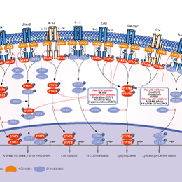
STAT阻害剤の選択性比較
Cell Data
| Cell Lines | Assay Type | Concentration | Incubation Time | 活性情報 | PMID |
|---|---|---|---|---|---|
| EHEB | Apoptosis Assay | 40 μM | 24 h | induces apoptosis | 23497075 |
| HEC50B | Apoptosis Assay | 20/100 μM | 24 h | induces apoptosis slightly | 23595697 |
| AN3CA | Apoptosis Assay | 20/100 μM | 24 h | induces apoptosis in a dose-dependent manner | 23595697 |
| HEC1A | Apoptosis Assay | 20/100 μM | 24 h | induces apoptosis in a dose-dependent manner | 23595697 |
| HEC50B | Growth Inhibition Assay | 100-500 μM | 24 h | inhibits cell growth slightly | 23595697 |
| AN3CA | Growth Inhibition Assay | 100-500 μM | 24 h | inhibits cell growth in a dose-dependent manner | 23595697 |
| HEC1A | Growth Inhibition Assay | 100-500 μM | 24 h | inhibits cell growth in a dose-dependent manner | 23595697 |
| Reh | Function Assay | 10 μM | 1/2/4 h | induces autophagy | 23681223 |
| Nalm-6 | Function Assay | 10 μM | 1/2/4 h | induces autophagy | 23681223 |
| DU-145 | Growth Inhibition Assay | 0-10 μg/ml | 48 h | inhibits cell growth in a dose-dependent manner | 23734815 |
| NALM6 | Apoptosis Assay | 5 μM | 24 h | induces cell apoptosis | 24057147 |
| I-83 | Apoptosis Assay | 5 μM | 24 h | induces cell apoptosis | 24057147 |
| BJAB | Apoptosis Assay | 5 μM | 24 h | induces cell apoptosis | 24057147 |
| HMECs | Function Assay | 100 μM | 36 h | inhibits IFNα mediated phosphorylation of STAT1 and STAT3, but not of STAT2 | 24211327 |
| HMECs | Function Assay | 100 μM | 36 h | inhibits IFNγ and LPS induced STAT1 phosphorylation and IRF1 expression | 24211327 |
| 624.38mel | Function Assay | 50-200 μM | 24 h | inhibits IDO activity independently of mRNA levels | 24911872 |
| MDA-231 | Function Assay | 50-200 μM | 24 h | inhibits IDO activity independently of mRNA levels | 24911872 |
| 624.38mel | Function Assay | 50 μM | 24 h | decreases IDO expression | 24911872 |
| MDA-231 | Function Assay | 100 μM | 24 h | decreases IDO expression | 24911872 |
| PBMC | Function Assay | 50/100 μM | 24 h | inhibits STAT1 phosphorylation | 24911872 |
| Raji | Function Assay | 3 μM | 24/48/72 h | induces accumulations of p53, p63 and p73 | 24940695 |
| KBM3/Bu2506 | Apoptosis Assay | 2.5 μM | 48 h | induces apoptosis slightly | 25111583 |
| MOLM 13 | Apoptosis Assay | 2.5 μM | 48 h | induces apoptosis slightly | 25111583 |
| THP-1 | Apoptosis Assay | 2.5 μM | 48 h | induces apoptosis slightly | 25111583 |
| MV-4-11 | Apoptosis Assay | 2.5 μM | 48 h | induces apoptosis slightly | 25111583 |
| Jeko-1 | Function Assay | 20 μM | 24 h | inhibits expression of IDO | 25940712 |
| CLL | Apoptosis Assay | 10 μM | 24-96 h | induces apoptotic cell death | 22207686 |
| MEC1 | Apoptosis Assay | 100 μM | 72 h | induces apoptosis significantly | 22132973 |
| U937 | Apoptosis Assay | 0.8 μM | 4-48 h | induces apoptosis slightly | 22074700 |
| U937 | Apoptosis Assay | 1 μM | 96 h | induces apoptosis slightly | 22023523 |
| Daudi | Apoptosis Assay | 20 μM | 96 h | induces apoptosis slightly | 22023523 |
| J45.01 | Apoptosis Assay | 1 μM | 96 h | induces apoptosis slightly | 22023523 |
| NALM-6 | Apoptosis Assay | 10 μM | 24 h | induces cell apoptosis slightly | 21699383 |
| JMV-3 | Apoptosis Assay | 10 μM | 24 h | induces cell apoptosis slightly | 21699383 |
| EHEB | Function Assay | 5-50 μM | 24 h | decreases p21 expression significantly | 21168391 |
| JVM-2 | Function Assay | 30 μM | 24 h | decreases p21 expression | 21168391 |
| KBM3/Bu2506 | Growth Inhibition Assay | 0.6 μM | 24 h | increases the cell fraction in S-phase | 20933509 |
| HLE-B3 | Function Assay | 25 μM | 48 h | blocks IFN-γ–induced STAT1 phosphorylation and IDO expression | 20435158 |
| SKW6.4 | Apoptosis Assay | 0.01-10 μM | 24/48 h | induces cell death in both time- and dose- dependent manner | 18092340 |
| CCRF-CEM | Function assay | 10 uM | 1 to 60 mins | Drug transport in human CCRF-CEM cells assessed as ENT1-mediated uptake at 10 uM after 1 to 60 mins by liquid scintillation counting analysis | 23388705 |
| K562 | Growth Inhibition Assay | 72 h | IC50=3.3 nM | 20307198 | |
| RPMI 8226 | Growth Inhibition Assay | 24 h | IC50=1.54 μM | 17976186 | |
| MM.1S | Growth Inhibition Assay | 48 h | IC50=13.48 μM | 17976186 | |
| MM.1R | Growth Inhibition Assay | 48 h | IC50=33.79 μM | 17976186 | |
| CLL5 | Antitumor assay | 48 hrs | Antitumor activity against CLL5 cells isolated from CLL patient assessed as cell viability after 48 hrs by FACS analysis, EC50 = 0.16 μM. | 24673739 | |
| K562 | Cytotoxicity assay | 72 hrs | Cytotoxicity against human paclitaxel-resistant K562 cells after 72 hrs by MTT assay, IC50 = 0.26 μM. | 20605656 | |
| CLL3 | Antitumor assay | 48 hrs | Antitumor activity against CLL3 cells isolated from CLL patient assessed as cell viability after 48 hrs by FACS analysis, EC50 = 0.35 μM. | 24673739 | |
| CLL4 | Antitumor assay | 48 hrs | Antitumor activity against CLL4 cells isolated from CLL patient assessed as cell viability after 48 hrs by FACS analysis, EC50 = 0.64 μM. | 24673739 | |
| CEM-DNR-B | Cytotoxicity assay | 72 hrs | Cytotoxicity against human CEM-DNR-B cells after 72 hrs by MTT assay, IC50 = 1.01 μM. | 20605656 | |
| CLL6 | Antitumor assay | 48 hrs | Antitumor activity against CLL6 cells isolated from CLL patient assessed as cell viability after 48 hrs by FACS analysis, EC50 = 1.6 μM. | 24673739 | |
| CLL2 | Antitumor assay | 48 hrs | Antitumor activity against CLL2 cells isolated from CLL patient assessed as cell viability after 48 hrs by FACS analysis, EC50 = 2.66 μM. | 24673739 | |
| HCT116 | Cytotoxicity assay | 72 hrs | Cytotoxicity against human HCT116 cells after 72 hrs by SRB assay, IC50 = 6.6 μM. | 25462277 | |
| PBMC | Cytotoxicity assay | 48 hrs | Cytotoxicity against patient PBMC after 48 hrs by CellTitre-Blue assay in presenc of mouse M210B4 cells, IC50 = 10 μM. | 25562417 | |
| CLL1 | Antitumor assay | 48 hrs | Antitumor activity against CLL1 cells isolated from CLL patient assessed as cell viability after 48 hrs by FACS analysis, EC50 = 17.1 μM. | 24673739 | |
| CEM | Cytotoxicity assay | 72 hrs | Cytotoxicity against human CEM cells after 72 hrs by MTT assay, IC50 = 19.49 μM. | 20605656 | |
| T47D | Cytotoxicity assay | 72 hrs | Cytotoxicity against human T47D cells after 72 hrs by SRB assay, IC50 = 46.2 μM. | 25462277 | |
| A549 | Cytotoxicity assay | 72 hrs | Cytotoxicity against human A549 cells after 72 hrs by MTT assay, IC50 = 47.44 μM. | 20605656 | |
| Reh | Growth Inhibition Assay | IC50 ∼10 μM | 23681223 | ||
| Nalm-6 | Growth Inhibition Assay | IC50 ∼10 μM | 23681223 | ||
| Nalm-6-FluR | Growth Inhibition Assay | IC50=250 μM | 25061101 | ||
| Molt-4 | Growth Inhibition Assay | IC50=180 μM | 25061101 | ||
| RPMI-8226 | Growth Inhibition Assay | IC50=500 μM | 25061101 | ||
| Mec-1 | Growth Inhibition Assay | IC50>500 μM | 25061101 | ||
| U2937 | Growth Inhibition Assay | IC50=16 μM | 25061101 | ||
| Reh | Growth Inhibition Assay | IC50=30 μM | 25061101 | ||
| Nalm-6 | Growth Inhibition Assay | IC50=18 μM | 25061101 | ||
| A549 | Growth Inhibition Assay | IC50=15.7±2.8 µM | 23377192 | ||
| A549 GAPDH-deficient | Growth Inhibition Assay | IC50=18.5±2.3 µM | 23377192 | ||
| RPMI 8226 | Growth Inhibition Assay | IC50=25.9 ± 3.7 μM | 21948264 | ||
| CEM | Growth Inhibition Assay | IC50=2.4 ± 0.4 μM | 21948264 | ||
| Raji | Growth Inhibition Assay | IC50=0.47 ± 0.04 μM | 21948264 | ||
| U937 | Growth Inhibition Assay | IC50=0.24 ± 0.04 μM | 21948264 | ||
| K562 | Growth Inhibition Assay | IC50=0.44 ± 0.05 μM | 21948264 | ||
| KBM3/Bu2506 | Growth Inhibition Assay | IC20=0.67 µM | 20933509 | ||
| MDA-MB-231 | Growth Inhibition Assay | IC50=4.0 μM | 20447390 | ||
| MCF-7 | Growth Inhibition Assay | IC50=15.0 μM | 20447390 | ||
| BW-225 | Growth Inhibition Assay | IC20=1.37 ×10⁻⁸ μM | 18661380 | ||
| OH-65 | Growth Inhibition Assay | IC20=1.37 ×10⁻⁸ μM | 18661380 | ||
| GR-145 | Growth Inhibition Assay | IC20=2.74 × 10⁻⁸ μM | 18661380 | ||
| A549 | Growth Inhibition Assay | IC20=5.48 × 10⁻⁸ μM | 18661380 | ||
| CaSki | Growth Inhibition Assay | IC20=1.37 × 10⁻⁷ μM | 18661380 | ||
| ZMK-1 | Growth Inhibition Assay | IC20=1.37 × 10⁻⁶ μM | 18661380 | ||
| U937 | Growth Inhibition Assay | IC50=3,200 ± 560 nM | 15930361 | ||
| primary CLL | Cytotoxicity assay | Cytotoxicity against human primary CLL cells, LD50 = 1.1 μM. | 25148392 | ||
| CHO | Function assay | Binding affinity determined by displacement of specific binding of [125I]N-(4-amino-3-iodophenethyl)-adenosine in membranes of CHO cells stably transfected with the rat adenosine A3 receptor, Ki = 10.4 μM. | 7707320 | ||
| JVM2 | Antitumor assay | Antitumor activity against human JVM2 cells assessed as cell viability after 48 hrs by FACS analysis, EC50 = 10.4 μM. | 24673739 | ||
| HeLa | Antitumor assay | Antitumor activity against human HeLa cells assessed as cell viability by MTT assay, EC50 = 16 μM. | 24673739 | ||
| TC32 | qHTS assay | qHTS of pediatric cancer cell lines to identify multiple opportunities for drug repurposing: Primary screen for TC32 cells | 29435139 | ||
| U-2 OS | qHTS assay | qHTS of pediatric cancer cell lines to identify multiple opportunities for drug repurposing: Primary screen for U-2 OS cells | 29435139 | ||
| A673 | qHTS assay | qHTS of pediatric cancer cell lines to identify multiple opportunities for drug repurposing: Primary screen for A673 cells | 29435139 | ||
| DAOY | qHTS assay | qHTS of pediatric cancer cell lines to identify multiple opportunities for drug repurposing: Primary screen for DAOY cells | 29435139 | ||
| Saos-2 | qHTS assay | qHTS of pediatric cancer cell lines to identify multiple opportunities for drug repurposing: Primary screen for Saos-2 cells | 29435139 | ||
| BT-37 | qHTS assay | qHTS of pediatric cancer cell lines to identify multiple opportunities for drug repurposing: Primary screen for BT-37 cells | 29435139 | ||
| RD | qHTS assay | qHTS of pediatric cancer cell lines to identify multiple opportunities for drug repurposing: Primary screen for RD cells | 29435139 | ||
| SK-N-SH | qHTS assay | qHTS of pediatric cancer cell lines to identify multiple opportunities for drug repurposing: Primary screen for SK-N-SH cells | 29435139 | ||
| MG 63 (6-TG R) | qHTS assay | qHTS of pediatric cancer cell lines to identify multiple opportunities for drug repurposing: Primary screen for MG 63 (6-TG R) cells | 29435139 | ||
| BT-12 | qHTS assay | qHTS of pediatric cancer cell lines to identify multiple opportunities for drug repurposing: Primary screen for BT-12 cells | 29435139 | ||
| NB1643 | qHTS assay | qHTS of pediatric cancer cell lines to identify multiple opportunities for drug repurposing: Primary screen for NB1643 cells | 29435139 | ||
| OHS-50 | qHTS assay | qHTS of pediatric cancer cell lines to identify multiple opportunities for drug repurposing: Primary screen for OHS-50 cells | 29435139 | ||
| BT-12 | qHTS assay | qHTS of pediatric cancer cell lines to identify multiple opportunities for drug repurposing: Confirmatory screen for BT-12 cells | 29435139 | ||
| LAN-5 | qHTS assay | qHTS of pediatric cancer cell lines to identify multiple opportunities for drug repurposing: Confirmatory screen for LAN-5 cells | 29435139 | ||
| NB-EBc1 | qHTS assay | qHTS of pediatric cancer cell lines to identify multiple opportunities for drug repurposing: Confirmatory screen for NB-EBc1 cells | 29435139 | ||
| SK-N-SH | qHTS assay | qHTS of pediatric cancer cell lines to identify multiple opportunities for drug repurposing: Confirmatory screen for SK-N-SH cells | 29435139 | ||
| Rh41 | qHTS assay | qHTS of pediatric cancer cell lines to identify multiple opportunities for drug repurposing: Primary screen for Rh41 cells | 29435139 | ||
| A673 | qHTS assay | qHTS of pediatric cancer cell lines to identify multiple opportunities for drug repurposing: Confirmatory screen for A673 cells) | 29435139 | ||
| BT-37 | qHTS assay | qHTS of pediatric cancer cell lines to identify multiple opportunities for drug repurposing: Confirmatory screen for BT-37 cells | 29435139 | ||
| MG 63 (6-TG R) | qHTS assay | qHTS of pediatric cancer cell lines to identify multiple opportunities for drug repurposing: Confirmatory screen for MG 63 (6-TG R) cells | 29435139 | ||
| Rh30 | qHTS assay | qHTS of pediatric cancer cell lines to identify multiple opportunities for drug repurposing: Confirmatory screen for Rh30 cells | 29435139 | ||
| U-2 OS | qHTS assay | qHTS of pediatric cancer cell lines to identify multiple opportunities for drug repurposing: Confirmatory screen for U-2 OS cells | 29435139 | ||
| OHS-50 | qHTS assay | qHTS of pediatric cancer cell lines to identify multiple opportunities for drug repurposing: Confirmatory screen for OHS-50 cells | 29435139 | ||
| SK-N-SH | qHTS assay | qHTS of pediatric cancer cell lines to identify multiple opportunities for drug repurposing: Orthogonal 3D viability screen for SK-N-SH cells | 29435139 | ||
| RD | qHTS assay | qHTS of pediatric cancer cell lines to identify multiple opportunities for drug repurposing: Confirmatory screen for RD cells | 29435139 | ||
| Daoy | qHTS assay | qHTS of pediatric cancer cell lines to identify multiple opportunities for drug repurposing: Orthogonal 3D viability screen for Daoy cells | 29435139 | ||
| TC32 | qHTS assay | qHTS of pediatric cancer cell lines to identify multiple opportunities for drug repurposing: Orthogonal 3D caspase screen for TC32 cells | 29435139 | ||
| TC32 | qHTS assay | qHTS of pediatric cancer cell lines to identify multiple opportunities for drug repurposing: Orthogonal 3D viability screen for TC32 cells | 29435139 | ||
| MG 63 (6-TG R) | qHTS assay | qHTS of pediatric cancer cell lines to identify multiple opportunities for drug repurposing: Orthogonal 3D viability screen for MG 63 (6-TG R) cells | 29435139 | ||
| SJ-GBM2 | qHTS assay | qHTS of pediatric cancer cell lines to identify multiple opportunities for drug repurposing: Primary screen for SJ-GBM2 cells | 29435139 | ||
| SK-N-MC | qHTS assay | qHTS of pediatric cancer cell lines to identify multiple opportunities for drug repurposing: Primary screen for SK-N-MC cells | 29435139 | ||
| NB-EBc1 | qHTS assay | qHTS of pediatric cancer cell lines to identify multiple opportunities for drug repurposing: Primary screen for NB-EBc1 cells | 29435139 | ||
| LAN-5 | qHTS assay | qHTS of pediatric cancer cell lines to identify multiple opportunities for drug repurposing: Primary screen for LAN-5 cells | 29435139 | ||
| Rh18 | qHTS assay | qHTS of pediatric cancer cell lines to identify multiple opportunities for drug repurposing: Primary screen for Rh18 cells | 29435139 | ||
| NB1643 | qHTS assay | qHTS of pediatric cancer cell lines to identify multiple opportunities for drug repurposing: Confirmatory screen for NB1643 cells | 29435139 | ||
| SK-N-MC | qHTS assay | qHTS of pediatric cancer cell lines to identify multiple opportunities for drug repurposing: Confirmatory screen for SK-N-MC cells | 29435139 | ||
| SJ-GBM2 | qHTS assay | qHTS of pediatric cancer cell lines to identify multiple opportunities for drug repurposing: Confirmatory screen for SJ-GBM2 cells | 29435139 | ||
| TC32 | qHTS assay | qHTS of pediatric cancer cell lines to identify multiple opportunities for drug repurposing: Confirmatory screen for TC32 cells | 29435139 | ||
| Rh18 | qHTS assay | qHTS of pediatric cancer cell lines to identify multiple opportunities for drug repurposing: Confirmatory screen for Rh18 cells | 29435139 | ||
| Saos-2 | qHTS assay | qHTS of pediatric cancer cell lines to identify multiple opportunities for drug repurposing: Confirmatory screen for Saos-2 cells | 29435139 | ||
| SJ-GBM2 | qHTS assay | qHTS of pediatric cancer cell lines to identify multiple opportunities for drug repurposing: Orthogonal 3D viability screen for SJ-GBM2 cells | 29435139 | ||
| RD | qHTS assay | qHTS of pediatric cancer cell lines to identify multiple opportunities for drug repurposing: Orthogonal 3D viability screen for RD cells | 29435139 | ||
| 他の多くの細胞株試験データをご覧になる場合はこちらをクリックして下さい | |||||
生物活性
| 製品説明 | Fludarabineは、STAT1 activation阻害剤であり、他のSTATsではなくSTAT1タンパク質(およびmRNA)の特異的な枯渇を引き起こします。また、血管平滑筋細胞におけるDNA synthesis阻害剤でもあります。Fludarabineはapoptosisを誘発します。 | |
|---|---|---|
| Targets |
|
| In Vitro | ||||
| In vitro |
Fludarabine efficiently inhibits the proliferation of RPMI 8226 cells with IC50 of 1.54 μg/mL. The IC50 of this compound against MM.1S and MM.1R cells is 13.48 μg/mL and 33.79 μg/mL, respectively. In contrast, U266 cells are resistant to this chemical with IC50 of 222.2 μg/mL. This compound treatment results in increased number of cells in the G1 phase of cell cycle, accompanied with a concomitant reduction of cells at the S phase of cell cycle in a time-dependent manner. It induces a cell cycle block and triggers apoptosis in MM cells. It triggers time-dependent cleavage of caspase-8, -9, and -3, -7, followed by PARP cleavage. It increases expression of Bax in a time-dependent fashion, while the expression of Bak doesn't change. After exposure to this chemical for 12 hours, RPMI 8226 cells shows a loss of membrane potential with 61.05% of the cells expressing low fluorescence of rhodamine 123 compared with 8.62% of cells in untreated control. To enhance solubility, it is formulated as the monophosphate (F-ara-AMP, fudarabine), which is instantaneously and quantitatively dephosphorylated to the parent nucleoside upon intravenous infusion. Inside the cells rephosphorylation occurs which leads to fuoroadenine arabinoside triphosphate (F-ara-ATP), the major cytotoxic metabolite of F-ara-A. It can also induce pro-inflammatory stimulation of monocytic cells, as evaluated by increased expression of ICAM-1 and IL-8 release. This compound does not affect the growth of ovarian cancer cell lines, whereas it induces marked and dose-dependent inhibition of proliferation in melanoma cell lines. It is an inhibitor of STAT1 that specifically reduces STAT1 without affecting other STAT family members. In addition to cytoplasmic accumulation, repeated low-dose cisplatin (RLDC) induces HMGB1 expression, which is marked suppressed by STAT1 knockdown. Consistently, this chemical suppresses HMGB1 expression during RLDC treatment dose-dependently in RLDC-treat renal tubular cells. |
|||
|---|---|---|---|---|
| 細胞実験 | 細胞株 | Dexamethasone-sensitive (MM.1S) and -resistant (MM.1R) human MM cell lines, RPMI8226 and U266 cell lines | ||
| 濃度 | 2 μg/mL | |||
| 反応時間 | 24 hours | |||
| 実験の流れ | After treated with Fludarabine or control, dexamethasone-sensitive (MM.1S) and -resistant (MM.1R) human MM cell lines, RPMI8226 and U266 cell lines (5 × 10⁵ cells) are washed twice in phosphate-buffered saline (PBS) and fixed with 70% ice-cold ethanol, then centrifuged and suspended in PBS containing 100 μg/mL RNase A. After incubated for 30 minutes at 37 ºC, samples are resuspended in 25 μg/mL propidium iodide. Flow cytometry is performed on a FACSCalibur automated system. Apoptosis is determined by Annexin V-FITC apoptosis detection kit, according to the manufacturer's instructions. For TUNEL (terminal deoxynucleotidyl transferase-mediated deoxyuridine triphosphate nick end labeling) assay, cells are analyzed by flow cytometry using the in situ cell death detection kit. |
|||
| 実験結果図 | Methods | Biomarkers | 結果図 | PMID |
| Western blot | procaspase-9 / procaspase-3 p-p53 / p53 STAT1 |
|
27223263 | |
| Immunofluorescence | α-SMA / Vimentin |
|
28322315 | |
| Growth inhibition assay | Cell viability |
|
24956101 | |
| In Vivo | ||
| In Vivo |
Tumors treated with PBS grow rapidly to approximately 10-fold their initial volume in 25 day, whereas, the tumors in the Fludarabine at 40 mg/kg increase less than 5-fold. A significant antitumor effect of 40 mg/kg of this compound on RPMI8226 tumor growth is demonstrated. RPMI8226 tumors treated with 40 mg/kg of this chemical at day 10 increase apoptotic nuclei. This compound is effective in suppressing RPMI8226 myeloma xenografts in SCID mice. |
|
|---|---|---|
| 動物実験 | 動物モデル | Severe combined immunodeficient (SCID) mice bearing RPMI 8226 cells |
| 投与量 | 40 mg/kg | |
| 投与経路 | Administered via i.p. | |
| NCT Number | Recruitment | Conditions | Sponsor/Collaborators | Start Date | Phases |
|---|---|---|---|---|---|
| NCT05390814 | Recruiting | Primary Central Nervous System Lymphoma |
Assistance Publique - Hôpitaux de Paris |
December 18 2023 | Phase 1 |
| NCT05201183 | Withdrawn | Acute Myeloid Leukemia|Chronic Myeloid Leukemia|Acute Lymphocytic Leukemia|Myelodysplastic Syndromes |
Naoyuki G. Saito M.D. Ph.D.|Indiana University |
October 2023 | Phase 1|Phase 2 |
| NCT05917405 | Recruiting | Acute Myeloid Leukemia in Remission |
Nantes University Hospital |
September 14 2023 | Phase 2 |
|
化学情報
| 分子量 | 285.23 | 化学式 | C10H12FN5O4 |
| CAS No. | 21679-14-1 | SDF | Download Fludarabine SDFをダウンロードする |
| Smiles | C1=NC2=C(N=C(N=C2N1C3C(C(C(O3)CO)O)O)F)N | ||
| 保管 | |||
|
In vitro |
DMSO : 57 mg/mL ( (199.83 mM); 吸湿したDMSOは溶解度を減少させます。新しいDMSOをご使用ください。) Water : Insoluble Ethanol : Insoluble |
モル濃度計算器 |
|
in vivo Add solvents to the product individually and in order. |
投与溶液組成計算機 | |||||
実験計算
投与溶液組成計算機(クリア溶液)
ステップ1:実験データを入力してください。(実験操作によるロスを考慮し、動物数を1匹分多くして計算・調製することを推奨します)
mg/kg
g
μL
匹
ステップ2:投与溶媒の組成を入力してください。(ロット毎に適した溶解組成が異なる場合があります。詳細については弊社までお問い合わせください)
% DMSO
%
% Tween 80
% ddH2O
%DMSO
%
計算結果:
投与溶媒濃度: mg/ml;
DMSOストック溶液調製方法: mg 試薬を μL DMSOに溶解する(濃度 mg/mL, 注:濃度が当該ロットのDMSO溶解度を超える場合はご連絡ください。 )
投与溶媒調製方法:Take μL DMSOストック溶液に μL PEG300,を加え、完全溶解後μL Tween 80,を加えて完全溶解させた後 μL ddH2O,を加え完全に溶解させます。
投与溶媒調製方法:μL DMSOストック溶液に μL Corn oil,を加え、完全溶解。
注意:1.ストック溶液に沈殿、混濁などがないことをご確認ください;
2.順番通りに溶剤を加えてください。次のステップに進む前に溶液に沈殿、混濁などがないことを確認してから加えてください。ボルテックス、ソニケーション、水浴加熱など物理的な方法で溶解を早めることは可能です。
技術サポート
ストックの作り方、阻害剤の保管方法、細胞実験や動物実験の際に注意すべき点など、製品を取扱う時に問い合わせが多かった質問に対しては取扱説明書でお答えしています。
他に質問がある場合は、お気軽にお問い合わせください。
* 必須
よくある質問(FAQ)
質問1:
how to re-suspend and deliver it for in vivo experiments?
回答
For S1491, we tested a vehicle: 30% Propylene glycol, 5% Tween 80, 65% D5W that you can resuspend this compound in at up to 30mg/ml. It is a suspension and can only be given via oral gavage.
納期 国内在庫品:受注日の翌日(15時までの受注分) *北海道、九州、沖縄への配送は受注日より2日以上 を要する場合あり 海外在庫品:受注後1〜2週間