
- 阻害剤
- 研究分野別
- PI3K/Akt/mTOR
- Epigenetics
- Methylation
- Immunology & Inflammation
- Protein Tyrosine Kinase
- Angiogenesis
- Apoptosis
- Autophagy
- ER stress & UPR
- JAK/STAT
- MAPK
- Cytoskeletal Signaling
- Cell Cycle
- TGF-beta/Smad
- 化合物ライブラリー
- 抗体
- 新製品
- お問い合わせ
ATF3 Antibody (Rabbit mAb) [B23E24]
Catalog No.: F1121
異なるサンプルでの発現パターンについては、“発現量と処理条件の例表 ”を参照してください。
Application:
Reactivity:
-
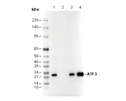 Lane 1: HeLa, Lane 2: HeLa (KO ATF3), Lane 3: HepG2, Lane 4: 293T
 -
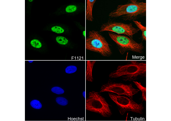 Immunofluorescent analysis of Hela cells using F1121 (green, 1:100), Hoechst (blue) and tubulin (Red).
カスタマーフィードバック(1)
キーポイント
WB
転写条件(ウェット): 200 mA, 60 min。
ATF3には2つのスプライスバリアントが存在し、二重バンドが現れる可能性があります(PMID:8622660)。
ATF3の基底発現は低いため、事前に誘導刺激を行うことを推奨します(PMID:19136462)。
転写条件(ウェット): 200 mA, 60 min。
ATF3には2つのスプライスバリアントが存在し、二重バンドが現れる可能性があります(PMID:8622660)。
ATF3の基底発現は低いため、事前に誘導刺激を行うことを推奨します(PMID:19136462)。
よくある問題とその対策

Fig. 1: WB (A375 Lysate) shows no bands (cells were not stimulated)
This antibody recognizes the ATF3 protein. In some cell lines, the endogenous expression level of ATF3 is low, and without stimulation, clear bands are not visible. Additionally, the RNA expression level of ATF3 in A375 cells is very low. Therefore, it is recommended to stimulate the cells during sample preparation, and for A375 cell lysates, increasing the sample loading amount is also recommended.
使用情報
| Dilution |
|---|
|
| Application |
|---|
| WB, IP, IF, ChIP |
| Source |
|---|
| Rabbit Monoclonal Antibody |
| Reactivity |
|---|
| Human, Mouse |
| Storage Buffer |
|---|
| PBS, pH 7.2+50% Glycerol+0.05% BSA+0.01% NaN₃ |
| Storage (from the date of receipt) |
|---|
| –20°C (avoid freeze-thaw cycles), 2 years |
| Predicted MW Observed MW |
|---|
| 21 kDa 21 kDa |
| *なぜ予測分子量と実際の分子量が異なるのか? 下記の原因により、実際の分子量が予測と異なる:タンパク質の翻訳後修飾(リン酸化/糖鎖付加),スプライシングバリアント,イソフォーム,相対的な電荷,ポリマー。 |
| ポジティブコントロール | Hap1; HeLa; A549; HCT116; HepG2; 293T; HEK-293; LnCap; THP-1 (treated with 80nM TPA overnight, then treated with 1 µg/mL LPS for 8 h) (PMID: 24973221); RAW 264.7 (treated with 1 µg/mL LPS for 2 h)(PMID: 24973221, PMID: 19136462) |
|---|---|
| ネガティブコントロール | Human liver; Mouse liver; Mouse heart; Jurkat; A431; THP-1; RAW 264.7; MEF |
サンプル処理データの例
| サンプル | 処理状況 |
| LX-2 | Medium Expression |
| PC-9 | Low Expression |
| クリックして、さらに多くのサンプルデータを表示 | |
*異なるヒト由来細胞や組織における発現量の予測については、以下をご参照ください: http://www.proteinatlas.org
プロトコール
| WB |
|---|
Western Blotting
Sample preparation
1. Aspirate media from cultures and Wash the cells with 1X PBS.
2. Lyse cells by adding 1X SDS sample buffer and transfer the extract to a microcentrifuge tube. Keep onice.
3. Sonicate for 10–15 sec to complete cell lysis and shear DNA.
4. Heat a 20 µl sample to 95–100°C for 5 min, then cool on ice.
5. Centrifuge for 5 min (with Microcentrifuge).
6. Load appropriate volumes of samples onto SDS-PAGE gel (loading quantity of protein sample depends on the concentration of extracted proteins).
NOTE: At the same time, please load the pre-stained molecular weight markers to determine molecular weights and verify electrotransfer.
7. Electrotransfer to nitrocellulose/PVDF membrane.
Membrane Blocking and Antibody Incubations
a. Blocking
1. (Optional) After transfer, wash the transferred membrane with TBS for 5 min at room temperature.
2. Incubate the membrane in the blocking buffer for 1 hr at room temperature.
3. Wash three times for 5 min each with TBST.
b. Antibodies Incubation
1. Incubate membrane and primary antibody (at the appropriate dilution and diluent recommended) in a primary antibody dilution buffer with gentle agitation overnight at 4°C.
2. Wash three times for 5 min each with TBST.
3. Incubate membrane with an appropriate second antibodydissolved in the blocking buffer with gentle agitation for 1 hr at room temperature.
4. Wash three times for 5 min each with TBST.
5. Proceed with detection.
Detection of Proteins
1. After antibodies incubation, Wash membrane three times for 5 minutes in TBST.
2. PrepareECL Reagent (or other chromogenic agents/substrate according to your second antibody). Mix well.
3. Incubate substrate with membrane for 1 minute, remove excess solution (membrane remains wet), wrap in plastic and expose in the imaging system. |
| WB |
|---|
Experimental Protocol:
Sample preparation
1. Tissue: Lyse the tissue sample by adding an appropriate volume of ice-cold RIPA/Nuclear Lysis Buffer (containing Protease Inhibitor Cocktail),and homogenize the tissue at a low temperature or lyse it by sonication on ice, then incubate on ice for 30 minutes. 2. Adherent cell: Aspirate the culture medium and transfer the cells into an EP tube. Wash the cells with ice-cold PBS twice. Add an appropriate volume of RIPA/Nuclear Lysis Buffer (containing Protease Inhibitor Cocktail), sonicate to lyse the cells, and incubate on ice for 30 minutes. 3. Suspension cell: Transfer the culture medium to a pre-cooled centrifuge tube. Centrifuge and aspirate the supernatant. Wash the cells with ice-cold PBS twice.Add an appropriate volume of RIPA/Nuclear Lysis Buffer (containing Protease Inhibitor Cocktail), sonicate to lyse the cells, and incubate on ice for 30 minutes. 4. Place the lysate into a pre-cooled microcentrifuge tube. Centrifuge at 4°C for 15 min. Collect the supernatant;
5. Remove a small volume of lysate to determine the protein concentration;
6. Combine the lysate with protein loading buffer. Boil 20 µL sample under 95-100°C for 5 min. Centrifuge for 5 min after cool down on ice.
Electrophoretic separation
1. According to the concentration of extracted protein, load appropriate amount of protein sample and marker onto SDS-PAGE gels for electrophoresis. Recommended separating gel (lower gel) concentration: 10%. Reference Table for Selecting SDS-PAGE Separation Gel Concentrations 2. Power up 80V for 30 minutes. Then the power supply is adjusted (110 V~150 V), the Marker is observed, and the electrophoresis can be stopped when the indicator band of the predyed protein Marker where the protein is located is properly separated. (Note that the current should not be too large when electrophoresis, too large current (more than 150 mA) will cause the temperature to rise, affecting the result of running glue. If high currents cannot be avoided, an ice bath can be used to cool the bath.)
Transfer membrane
1. Take out the converter, soak the clip and consumables in the pre-cooled converter;
2. Activate PVDF membrane with methanol for 1 min and rinse with transfer buffer;
3. Install it in the order of "black edge of clip - sponge - filter paper - filter paper - glue -PVDF membrane - filter paper - filter paper - sponge - white edge of clip"; 4. The protein was electrotransferred to PVDF membrane. ( 0.45 µm PVDF membrane is recommended ) Reference Table for Selecting PVDF Membrane Pore Size Specifications Recommended conditions for wet transfer: 200 mA, 60 min. ( Note that the transfer conditions can be adjusted according to the protein size. For high-molecular-weight proteins, a higher current and longer transfer time are recommended. However, ensure that the transfer tank remains at a low temperature to prevent gel melting.)
Block
1. After electrotransfer, wash the film with TBST at room temperature for 5 minutes;
2. Incubate the film in the blocking solution for 1 hour at room temperature;
3. Wash the film with TBST for 3 times, 5 minutes each time.
Antibody incubation
1. Use 5% skim milk powder to prepare the primary antibody working liquid (recommended dilution ratio for primary antibody 1:1000), gently shake and incubate with the film at 4°C overnight; 2. Wash the film with TBST 3 times, 5 minutes each time;
3. Add the secondary antibody to the blocking solution and incubate with the film gently at room temperature for 1 hour;
4. After incubation, wash the film with TBST 3 times for 5 minutes each time.
Antibody staining
553. Add the prepared ECL luminescent substrate (or select other color developing substrate according to the second antibody) and mix evenly;
2. Incubate with the film for 1 minute, remove excess substrate (keep the film moist), wrap with plastic film, and expose in the imaging system. |
| IF |
|---|
Experimental Protocol:
Sample Preparation
1. Adherent Cells: Place a clean, sterile coverslip in a culture dish. Once the cells grow to near confluence as a monolayer, remove the coverslip for further use.
2. Suspension Cells: Seed the cells onto a clean, sterile slide coated with poly-L-lysine.
3. Frozen Sections: Allow the slide to thaw at room temperature. Wash it with pure water or PBS for 2 times, 3 minutes each time.
4. Paraffin Sections: Deparaffinization and rehydration. Wash the slide with pure water or PBS for 3 times, 3 minutes each time. Then perform antigen retrieval.
Fixation
1. Fix the cell coverslips/spots or tissue sections at room temperature using a fixative such as 4% paraformaldehyde (4% PFA) for 10-15 minutes.
2. Wash the sample with PBS for 3 times, 3 minutes each time.
Permeabilization
1.Add a detergent such as 0.1–0.3% Triton X-100 to the sample and incubate at room temperature for 10–20 minutes.
(Note: This step is only required for intracellular antigens. For antigens expressed on the cell membrane, this step is unnecessary.)
Wash the sample with PBS for 3 times, 3 minutes each time.
Blocking
Add blocking solution and incubate at room temperature for at least 1 hour. (Common blocking solutions include: serum from the same source as the secondary antibody, BSA, or goat serum.)
Note: Ensure the sample remains moist during and after the blocking step to prevent drying, which can lead to high background.
Immunofluorescence Staining (Day 1)
1. Remove the blocking solution and add the diluted primary antibody.
2. Incubate the sample in a humidified chamber at 4°C overnight.
Immunofluorescence Staining (Day 2)
1. Remove the primary antibody and wash with PBST for 3 times, 5 minutes each time.
2. Add the diluted fluorescent secondary antibody and incubate in the dark at 4°C for 1–2 hours.
3. Remove the secondary antibody and wash with PBST for 3 times, 5 minutes each time.
4. Add diluted DAPI and incubate at room temperature in the dark for 5–10 minutes.
5. Wash with PBST for 3 times, 5 minutes each time.
Mounting
1. Mount the sample with an anti-fade mounting medium.
2. Allow the slide to dry at room temperature overnight in the dark.
3. Store the slide in a slide storage box at 4°C, protected from light.
|
生物学的記述
| Specificity |
|---|
ATF3 Antibody (Rabbit mAb) [B23E24] detects endogenous levels of total ATF3 protein. Stimulation may be required to allow detection of the target protein due to low levels of endogenous expression in some samples. |
| タンパク質の局在 |
|---|
| 細胞核 |
| Uniprot ID |
|---|
| P18847 |
| Clone |
|---|
| B23E24 |
| Synonym(s) |
|---|
| ATF3 |
| Background |
|---|
Activating transcription factor 3 (ATF3) is a member of the ATF/CREB family, binding to the cyclic AMP response element (CRE) with the consensus sequence TGACGTCA. It is a stress-induced transcription factor that regulates metabolism, immunity, and oncogenesis, acting as a central hub in the cellular adaptive-response network. ATF3 is induced by various extracellular signals, including ER stress, cytokines, chemokines, and LPS. |
| References |
|---|
技術サポート
ストックの作り方、阻害剤の保管方法、細胞実験や動物実験の際に注意すべき点など、製品を取扱う時に問い合わせが多かった質問に対しては取扱説明書でお答えしています。
他に質問がある場合は、お気軽にお問い合わせください。
* 必須
納期 国内在庫品:受注日の翌日(15時までの受注分) *北海道、九州、沖縄への配送は受注日より2日以上 を要する場合あり 海外在庫品:受注後1〜2週間