
- 阻害剤
- 研究分野別
- PI3K/Akt/mTOR
- Epigenetics
- Methylation
- Immunology & Inflammation
- Protein Tyrosine Kinase
- Angiogenesis
- Apoptosis
- Autophagy
- ER stress & UPR
- JAK/STAT
- MAPK
- Cytoskeletal Signaling
- Cell Cycle
- TGF-beta/Smad
- 化合物ライブラリー
- 抗体
- 新製品
- お問い合わせ
c-Fos Antibody [M9L15]
Catalog No.: F0267
Application:
Reactivity:
-
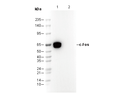 Lane 1: HeLa (serum-starved overnight; TPA,4 hours) , Lane 2: HeLa (serum-starved overnight)
カスタマーフィードバック(1)
キーポイント
WB
Helaなどの特定の細胞株では、c-Fosの内因性レベルが比較的低いです。高い内因性c-Fosレベルを維持するために、誘導刺激(一晩の血清飢餓処理とさらに4時間のTPA処理など)を加えることが推奨されます。
刺激後、c-Fosの発現は急速かつ短命です。タンパク質の分解を防ぐため、刺激終了後は直ちにサンプルを採取することが推奨されます。
よくある問題とその対策
Fig. 1: WB (Human Pancreatic Cancer Cell Lysate) shows no bands (cells were not stimulated)
This antibody recognizes the c-Fos protein. In some cell lines, the endogenous expression level of c-Fos is low, requiring stimulation to see clear bands. It is recommended to perform overnight serum starvation followed by TPA treatment for stimulation. The expression of c-Fos is rapid and transient after stimulation, so the samples should be collected immediately after stimulation to prevent protein degradation.
使用情報
| Dilution |
|---|
|
| Application |
|---|
| WB, FCM, ChIP |
| Source |
|---|
| Rabbit Monoclonal Antibody |
| Reactivity |
|---|
| Human, Mouse, Rat |
| Storage Buffer |
|---|
| PBS, pH 7.2+50% Glycerol+0.05% BSA+0.01% NaN₃ |
| Storage (from the date of receipt) |
|---|
| –20°C (avoid freeze-thaw cycles), 2 years |
| Predicted MW |
|---|
| 62 kDa |
| ポジティブコントロール | mouse cerebral cortex; mouse pons; Hela (serum-starved overnight + 400nM TPA-stimulated, 4 h); H-4-II-E (serum-starved overnight + 400nM TPA-stimulated, 4 h); NIH/3T3 (serum-starved overnight + 400nM TPA-stimulated, 4 h); PC-12 (serum-starved overnight, treated with h-βNGF, 50ng/ml, 2h) |
|---|---|
| ネガティブコントロール | Hela; H-4-II-E; NIH/3T3; PC-12 |
サンプル処理データの例
| サンプル | 処理状況 |
| Hela | Serum-starvation (overnight) + TPA (400nM, 4 h) |
| H-4-II-E | Serum-starvation (overnight) + TPA (400nM, 4 h) |
| NIH/3T3 | Serum-starvation (overnight) + TPA (400nM, 4 h) |
| クリックして、さらに多くのサンプルデータを表示 | |
*異なるヒト由来細胞や組織における発現量の予測については、以下をご参照ください: http://www.proteinatlas.org
プロトコール
| WB |
|---|
Experimental Protocol:
Sample preparation
1. Tissue: Lyse the tissue sample by adding an appropriate volume of ice-cold RIPA/NP-40 Lysis Buffer (containing Protease Inhibitor Cocktail),and homogenize the tissue at a low temperature or lyse it by sonication on ice, then incubate on ice for 30 minutes. 2. Adherent cell: Aspirate the culture medium and transfer the cells into an EP tube. Wash the cells with ice-cold PBS twice. Add an appropriate volume of RIPA/NP-40 Lysis Buffer (containing Protease Inhibitor Cocktail), sonicate to lyse the cells, and incubate on ice for 30 minutes. 3. Suspension cell: Transfer the culture medium to a pre-cooled centrifuge tube. Centrifuge and aspirate the supernatant. Wash the cells with ice-cold PBS twice.Add an appropriate volume of RIPA/NP-40 Lysis Buffer (containing Protease Inhibitor Cocktail), sonicate to lyse the cells, and incubate on ice for 30 minutes. 4. Place the lysate into a pre-cooled microcentrifuge tube. Centrifuge at 4°C for 15 min. Collect the supernatant;
5. Remove a small volume of lysate to determine the protein concentration;
6. Combine the lysate with protein loading buffer. Boil 20 µL sample under 95-100°C for 5 min. Centrifuge for 5 min after cool down on ice.
Electrophoretic separation
1. According to the concentration of extracted protein, load appropriate amount of protein sample and marker onto SDS-PAGE gels for electrophoresis. Recommended separating gel (lower gel) concentration: 10%. Reference Table for Selecting SDS-PAGE Separation Gel Concentrations 2. Power up 80V for 30 minutes. Then the power supply is adjusted (110 V~150 V), the Marker is observed, and the electrophoresis can be stopped when the indicator band of the predyed protein Marker where the protein is located is properly separated. (Note that the current should not be too large when electrophoresis, too large current (more than 150 mA) will cause the temperature to rise, affecting the result of running glue. If high currents cannot be avoided, an ice bath can be used to cool the bath.)
Transfer membrane
1. Take out the converter, soak the clip and consumables in the pre-cooled converter;
2. Activate PVDF membrane with methanol for 1 min and rinse with transfer buffer;
3. Install it in the order of "black edge of clip - sponge - filter paper - filter paper - glue -PVDF membrane - filter paper - filter paper - sponge - white edge of clip"; 4. The protein was electrotransferred to PVDF membrane. ( 0.45 µm PVDF membrane is recommended ) Reference Table for Selecting PVDF Membrane Pore Size Specifications Recommended conditions for wet transfer: 200 mA, 120 min. ( Note that the transfer conditions can be adjusted according to the protein size. For high-molecular-weight proteins, a higher current and longer transfer time are recommended. However, ensure that the transfer tank remains at a low temperature to prevent gel melting.)
Block
1. After electrotransfer, wash the film with TBST at room temperature for 5 minutes;
2. Incubate the film in the blocking solution for 1 hour at room temperature;
3. Wash the film with TBST for 3 times, 5 minutes each time.
Antibody incubation
1. Use 5% skim milk powder to prepare the primary antibody working liquid (recommended dilution ratio for primary antibody 1:1000), gently shake and incubate with the film at 4°C overnight; 2. Wash the film with TBST 3 times, 5 minutes each time;
3. Add the secondary antibody to the blocking solution and incubate with the film gently at room temperature for 1 hour;
4. After incubation, wash the film with TBST 3 times for 5 minutes each time.
Antibody staining
928. Add the prepared ECL luminescent substrate (or select other color developing substrate according to the second antibody) and mix evenly;
2. Incubate with the film for 1 minute, remove excess substrate (keep the film moist), wrap with plastic film, and expose in the imaging system. |
生物学的記述
| Specificity |
|---|
c-Fos Antibody [M9L15] detects endogenous levels of total c-Fos protein. |
| タンパク質の局在 |
|---|
| 細胞質、小胞体、細胞核 |
| Uniprot ID |
|---|
| P01100 |
| Clone |
|---|
| M9L15 |
| Background |
|---|
The Fos family of nuclear oncogenes consists of c-Fos, FosB, Fos-related antigen 1 (FRA1), and Fos-related antigen 2 (FRA2). Unlike other Fos proteins, which exist as a single isoform, FosB has two variants: the full-length FosB and a truncated version known as FosB2 (Delta FosB), which lacks the carboxy-terminal 101 amino acids. Fos proteins are rapidly and transiently expressed in response to various extracellular signals such as growth factors, cytokines, neurotransmitters, polypeptide hormones, and stress. These proteins dimerize with Jun family members (c-Jun, JunB, and JunD) to form the transcription factor Activator Protein-1 (AP-1), which binds to TRE/AP-1 elements in DNA to regulate gene transcription. Fos and Jun proteins have a leucine-zipper motif essential for dimerization and a basic domain that facilitates DNA binding. The different Fos/Jun heterodimers have varying capacities to activate AP-1 dependent genes. Beyond expression levels, phosphorylation of Fos proteins by Erk kinases in response to external stimuli can enhance their transcriptional activity. Specifically, phosphorylation of c-Fos at Ser32 and Thr232 by Erk5 increases its stability and nuclear localization. Abnormal expression of c-Fos, FosB, or FRA2 can lead to neoplastic transformation of cells. |
| References |
|---|
技術サポート
ストックの作り方、阻害剤の保管方法、細胞実験や動物実験の際に注意すべき点など、製品を取扱う時に問い合わせが多かった質問に対しては取扱説明書でお答えしています。
他に質問がある場合は、お気軽にお問い合わせください。
* 必須
納期 国内在庫品:受注日の翌日(15時までの受注分) *北海道、九州、沖縄への配送は受注日より2日以上 を要する場合あり 海外在庫品:受注後1〜2週間