
- 阻害剤
- 研究分野別
- PI3K/Akt/mTOR
- Epigenetics
- Methylation
- Immunology & Inflammation
- Protein Tyrosine Kinase
- Angiogenesis
- Apoptosis
- Autophagy
- ER stress & UPR
- JAK/STAT
- MAPK
- Cytoskeletal Signaling
- Cell Cycle
- TGF-beta/Smad
- 化合物ライブラリー
- 抗体
- 新製品
- お問い合わせ
CaSR Antibody (Mouse mAb) [A19J6]
Catalog No.: F1579
Application:
Reactivity:
-
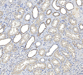 Immunohistochemical analysis of formalin fixed paraffin embedded human kidney tissue with F1579 at 1:100 dilution.
当該製品は品切れ状态で、メールアドレスをご教示いただければ、お客様に返信いたします。
代表番号: 045-509-1970|電子メール:sales@selleck.co.jp
使用情報
| Dilution |
|---|
|
| Application |
|---|
| IHC, FCM |
| Source |
|---|
| Mouse Monoclonal Antibody |
| Reactivity |
|---|
| Mouse, Human |
| Storage Buffer |
|---|
| PBS, pH 7.2+50% Glycerol+0.05% BSA+0.01% NaN3 |
| Storage (from the date of receipt) |
|---|
| -20°C (avoid freeze-thaw cycles), 2 years |
| ポジティブコントロール | Rat brain cerebral cortex tissue; Mouse stomach tissue; Human brain tissue; Human kidney tissue; SH-SY5Y cells |
|---|---|
| ネガティブコントロール |
プロトコール
| IHC |
|---|
Experimental Protocol:
Deparaffinization/Rehydration
1. Deparaffinize/hydrate sections:
2. Incubate sections in three washes of xylene for 5 min each.
3. Incubate sections in two washes of 100% ethanol for 10 min each.
4. Incubate sections in two washes of 95% ethanol for 10 min each.
5. Wash sections two times in dH2O for 5 min each.
6.Antigen retrieval: For Citrate: Heat slides in a microwave submersed in 1X citrate unmasking solution until boiling is initiated; continue with 10 min at a sub-boiling temperature (95°-98°C). Cool slides on bench top for 30 min.
Staining
1. Wash sections in dH2O three times for 5 min each.
2. Incubate sections in 3% hydrogen peroxide for 10 min.
3. Wash sections in dH2O two times for 5 min each.
4. Wash sections in wash buffer for 5 min.
5. Block each section with 100–400 µl of blocking solution for 1 hr at room temperature.
6. Remove blocking solution and add 100–400 µl primary antibody diluent in to each section. Incubate overnight at 4°C.
7. Remove antibody solution and wash sections with wash buffer three times for 5 min each.
8. Cover section with 1–3 drops HRPas needed. Incubate in a humidified chamber for 30 min at room temperature.
9. Wash sections three times with wash buffer for 5 min each.
10. Add DAB Chromogen Concentrate to DAB Diluent and mix well before use.
11. Apply 100–400 µl DAB to each section and monitor closely. 1–10 min generally provides an acceptable staining intensity.
12. Immerse slides in dH2O.
13. If desired, counterstain sections with hematoxylin.
14. Wash sections in dH2O two times for 5 min each.
15. Dehydrate sections: Incubate sections in 95% ethanol two times for 10 sec each; Repeat in 100% ethanol, incubating sections two times for 10 sec each; Repeat in xylene, incubating sections two times for 10 sec each.
16. Mount sections with coverslips and mounting medium.
|
生物学的記述
| Specificity |
|---|
| CaSR Antibody (Mouse mAb) [A19J6] detects endogenous levels of total CaSR protein. |
| タンパク質の局在 |
|---|
| 細胞膜、細胞内膜系 |
| Uniprot ID |
|---|
| P41180 |
| Clone |
|---|
| A19J6 |
| Synonym(s) |
|---|
| GPRC2A; PCAR1; CASR; Extracellular calcium-sensing receptor; CaR; CaSR; hCasR; Parathyroid cell calcium-sensing receptor 1; PCaR1 |
| Background |
|---|
| The calcium-sensing receptor (CaSR) is a class C G protein–coupled receptor that functions as an extracellular calcium sensor, translating changes in ionized calcium concentration into coordinated endocrine and renal responses. It contains a large extracellular Venus flytrap domain that binds calcium and other polycations, a cysteine‑rich region, and a seven‑transmembrane domain that couples predominantly to Gq/11 and Gi/o proteins. Activation of Gq/11 triggers phospholipase Cβ, leading to inositol trisphosphate–mediated calcium mobilization and diacylglycerol–dependent activation of protein kinase C, while Gi/o coupling suppresses adenylate cyclase and lowers cAMP levels. In parathyroid chief cells, increased extracellular calcium engages CaSR to inhibit parathyroid hormone (PTH) secretion and promote PTH internalization, establishing a major negative feedback loop for systemic calcium homeostasis. In the kidney, CaSR signaling in the thick ascending limb and collecting duct influences calcium and magnesium reabsorption, phosphate handling, and urine concentration by interacting with transporters such as NKCC2, ROMK, and ENaC, and by intersecting with MAPK and mTOR pathways that modulate cell growth and tubular function. CaSR‑dependent signaling also contributes to regulation of vitamin D metabolism and bone turnover through its effects on PTH and downstream endocrine axes. Germline loss‑of‑function mutations in CaSR cause familial hypocalciuric hypercalcemia and neonatal severe hyperparathyroidism, characterized by elevated serum calcium and inappropriately normal or high PTH, whereas gain‑of‑function mutations lead to autosomal dominant hypocalcemia with low calcium and suppressed PTH. |
| References |
|---|
技術サポート
ストックの作り方、阻害剤の保管方法、細胞実験や動物実験の際に注意すべき点など、製品を取扱う時に問い合わせが多かった質問に対しては取扱説明書でお答えしています。
他に質問がある場合は、お気軽にお問い合わせください。
* 必須
納期 国内在庫品:受注日の翌日(15時までの受注分) *北海道、九州、沖縄への配送は受注日より2日以上 を要する場合あり 海外在庫品:受注後1〜2週間