
- 阻害剤
- 研究分野別
- PI3K/Akt/mTOR
- Epigenetics
- Methylation
- Immunology & Inflammation
- Protein Tyrosine Kinase
- Angiogenesis
- Apoptosis
- Autophagy
- ER stress & UPR
- JAK/STAT
- MAPK
- Cytoskeletal Signaling
- Cell Cycle
- TGF-beta/Smad
- 化合物ライブラリー
- 抗体
- 新製品
- お問い合わせ
LYVE1 Antibody [P8K5]
Catalog No.: F3736
Application:
Reactivity:
-
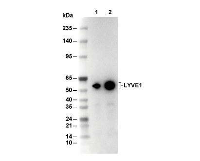 Lane 1: Jurkat, Lane 2: K562
当該製品は品切れ状态で、メールアドレスをご教示いただければ、お客様に返信いたします。
代表番号: 045-509-1970|電子メール:sales@selleck.co.jp
キーポイント
WB
転写条件(ウェット): 200 mA, 60 min。
90秒以上の露光(暴露)を推奨します。
転写条件(ウェット): 200 mA, 60 min。
90秒以上の露光(暴露)を推奨します。
使用情報
| Dilution |
|---|
|
| Application |
|---|
| WB, IP, IHC |
| Source |
|---|
| Rabbit Monoclonal Antibody |
| Reactivity |
|---|
| Human |
| Storage Buffer |
|---|
| PBS, pH 7.2+50% Glycerol+0.05% BSA+0.01% NaN3 |
| Storage (from the date of receipt) |
|---|
| -20°C (avoid freeze-thaw cycles), 2 years |
| Predicted MW Observed MW |
|---|
| 35 kDa 50-70 kDa |
| *なぜ予測分子量と実際の分子量が異なるのか? 下記の原因により、実際の分子量が予測と異なる:タンパク質の翻訳後修飾(リン酸化/糖鎖付加),スプライシングバリアント,イソフォーム,相対的な電荷,ポリマー。 |
| ポジティブコントロール | Human fetal lung tissue; Human liver tissue; Human lymph node tissue; Human tonsil tissue; Human fetal spleen tissue; Human colon tissue |
|---|---|
| ネガティブコントロール | Human brain tissue |
プロトコール
| WB |
|---|
Experimental Protocol:
Sample preparation
1. Tissue: Lyse the tissue sample by adding an appropriate volume of ice-cold RIPA/NP-40 Lysis Buffer (containing Protease Inhibitor Cocktail),and homogenize the tissue at a low temperature. 2. Adherent cell: Aspirate the culture medium and wash the cells with ice-cold PBS twice. Lyse the cells by adding an appropriate volume of RIPA/NP-40 Lysis Buffer (containing Protease Inhibitor Cocktail) and put the sample on ice for 5 min. 3. Suspension cell: Transfer the culture medium to a pre-cooled centrifuge tube. Centrifuge and aspirate the supernatant. Wash the cells with ice-cold PBS twice. Lyse the cells by adding an appropriate volume of RIPA/NP-40 Lysis Buffer (containing Protease Inhibitor Cocktail) and put the sample on ice for 5 min. 4. Place the lysate into a pre-cooled microcentrifuge tube. Centrifuge at 4°C for 15 min. Collect the supernatant;
5. Remove a small volume of lysate to determine the protein concentration;
6. Combine the lysate with protein loading buffer. Boil 20 µL sample under 95-100°C for 5 min. Centrifuge for 5 min after cool down on ice.
Electrophoretic separation
1. According to the concentration of extracted protein, load appropriate amount of protein sample and marker onto SDS-PAGE gels for electrophoresis. Recommended separating gel (lower gel) concentration: 10%. Reference Table for Selecting SDS-PAGE Separation Gel Concentrations 2. Power up 80V for 30 minutes. Then the power supply is adjusted (110 V~150 V), the Marker is observed, and the electrophoresis can be stopped when the indicator band of the predyed protein Marker where the protein is located is properly separated. (Note that the current should not be too large when electrophoresis, too large current (more than 150 mA) will cause the temperature to rise, affecting the result of running glue. If high currents cannot be avoided, an ice bath can be used to cool the bath.)
Transfer membrane
1. Take out the converter, soak the clip and consumables in the pre-cooled converter;
2. Activate PVDF membrane with methanol for 1 min and rinse with transfer buffer;
3. Install it in the order of "black edge of clip - sponge - filter paper - filter paper - glue -PVDF membrane - filter paper - filter paper - sponge - white edge of clip"; 4. The protein was electrotransferred to PVDF membrane. ( 0.45 µm PVDF membrane is recommended ) Reference Table for Selecting PVDF Membrane Pore Size Specifications Recommended conditions for wet transfer: 200 mA, 60 min. ( Note that the transfer conditions can be adjusted according to the protein size. For high-molecular-weight proteins, a higher current and longer transfer time are recommended. However, ensure that the transfer tank remains at a low temperature to prevent gel melting.)
Block
1. After electrotransfer, wash the film with TBST at room temperature for 5 minutes;
2. Incubate the film in the blocking solution for 1 hour at room temperature;
3. Wash the film with TBST for 3 times, 5 minutes each time.
Antibody incubation
1. Use 5% skim milk powder to prepare the primary antibody working liquid (recommended dilution ratio for primary antibody 1:1000), gently shake and incubate with the film at 4°C overnight; 2. Wash the film with TBST 3 times, 5 minutes each time;
3. Add the secondary antibody to the blocking solution and incubate with the film gently at room temperature for 1 hour;
4. After incubation, wash the film with TBST 3 times for 5 minutes each time.
Antibody staining
1. Add the prepared ECL luminescent substrate (or select other color developing substrate according to the second antibody) and mix evenly;
2. Incubate with the film for 1 minute, remove excess substrate (keep the film moist), wrap with plastic film, and expose in the imaging system. (Exposure time of at least 120s is recommended) |
| IHC |
|---|
Experimental Protocol:
Deparaffinization/Rehydration
1. Deparaffinize/hydrate sections:
2. Incubate sections in three washes of xylene for 5 min each.
3. Incubate sections in two washes of 100% ethanol for 10 min each.
4. Incubate sections in two washes of 95% ethanol for 10 min each.
5. Wash sections two times in dH2O for 5 min each.
6.Antigen retrieval: For Citrate: Heat slides in a microwave submersed in 1X citrate unmasking solution until boiling is initiated; continue with 10 min at a sub-boiling temperature (95°-98°C). Cool slides on bench top for 30 min.
Staining
1. Wash sections in dH2O three times for 5 min each.
2. Incubate sections in 3% hydrogen peroxide for 10 min.
3. Wash sections in dH2O two times for 5 min each.
4. Wash sections in wash buffer for 5 min.
5. Block each section with 100–400 µl of blocking solution for 1 hr at room temperature.
6. Remove blocking solution and add 100–400 µl primary antibody diluent in to each section. Incubate overnight at 4°C.
7. Remove antibody solution and wash sections with wash buffer three times for 5 min each.
8. Cover section with 1–3 drops HRPas needed. Incubate in a humidified chamber for 30 min at room temperature.
9. Wash sections three times with wash buffer for 5 min each.
10. Add DAB Chromogen Concentrate to DAB Diluent and mix well before use.
11. Apply 100–400 µl DAB to each section and monitor closely. 1–10 min generally provides an acceptable staining intensity.
12. Immerse slides in dH2O.
13. If desired, counterstain sections with hematoxylin.
14. Wash sections in dH2O two times for 5 min each.
15. Dehydrate sections: Incubate sections in 95% ethanol two times for 10 sec each; Repeat in 100% ethanol, incubating sections two times for 10 sec each; Repeat in xylene, incubating sections two times for 10 sec each.
16. Mount sections with coverslips and mounting medium.
|
生物学的記述
| Specificity |
|---|
| LYVE1 Antibody [P8K5] detects endogenous levels of total LYVE1 protein. |
| タンパク質の局在 |
|---|
| 細胞膜、細胞内膜系 |
| Uniprot ID |
|---|
| Q9Y5Y7 |
| Clone |
|---|
| P8K5 |
| Synonym(s) |
|---|
| CRSBP1; HAR; XLKD1; UNQ230/PRO263; LYVE1; Lymphatic vessel endothelial hyaluronic acid receptor 1; LYVE-1; Cell surface retention sequence-binding protein 1; Extracellular link domain-containing protein 1; Hyaluronic acid receptor; CRSBP-1 |
| Background |
|---|
| LYVE1 is a type I transmembrane glycoprotein and member of the Link module superfamily of hyaluronan (HA)-binding proteins, closely related to CD44, and serves as a highly specific marker of lymphatic endothelial cells. The 322-amino-acid protein features an extracellular N-terminal domain (residues 21–233) containing the signature HA-binding Link module (residues 48–162). This module forms a deep binding groove comprised of β-sheets and loops that capture HA chain ends through critical interactions involving Arg104, Lys107, Tyr86, and Trp115. The protein also has a single-span transmembrane helix (residues 234–255) and a short cytoplasmic tail (residues 256–322) equipped with dileucine motifs that mediate endocytosis. Unlike CD44, which binds immobilized HA with high affinity, LYVE1 preferentially recognizes soluble or pericellular HA on leukocyte glycocalyces. This interaction relies on a shear-resistant, end-on threading mechanism, where the non-reducing end of HA threads into the LYVE1 groove, requiring conformational changes like Arg104 flip and Lys107 shift to unblock the β4/β5 clasp. LYVE1 is particularly enriched at lymphatic blind-end junctions, where it facilitates dendritic cell (DC) and leukocyte transmigration by relaxing VE-cadherin junctions via Rac1/PI3K signaling. Soluble HA engagement also triggers VEGFR3-independent PI3K/AKT activation, promoting lymphangiogenesis during inflammation. LYVE1 mediates the internalization and lysosomal degradation of HA through clathrin-independent endocytosis, helping to clear interstitial HA, maintain tissue fluid balance, and prevent edema. In the tumor microenvironment, upregulation of LYVE1 on new lymphatic vessels enables binding to HA-rich metastasis clusters, enhancing VEGF-C-driven lymphatic sprouting and facilitating tumor spread to sentinel lymph nodes. LYVE1 deficiency impairs DC migration and T-cell priming, leading to reduced immune responses against pathogens. |
| References |
|---|
技術サポート
ストックの作り方、阻害剤の保管方法、細胞実験や動物実験の際に注意すべき点など、製品を取扱う時に問い合わせが多かった質問に対しては取扱説明書でお答えしています。
他に質問がある場合は、お気軽にお問い合わせください。
* 必須
納期 国内在庫品:受注日の翌日(15時までの受注分) *北海道、九州、沖縄への配送は受注日より2日以上 を要する場合あり 海外在庫品:受注後1〜2週間