
- 阻害剤
- 研究分野別
- PI3K/Akt/mTOR
- Epigenetics
- Methylation
- Immunology & Inflammation
- Protein Tyrosine Kinase
- Angiogenesis
- Apoptosis
- Autophagy
- ER stress & UPR
- JAK/STAT
- MAPK
- Cytoskeletal Signaling
- Cell Cycle
- TGF-beta/Smad
- 化合物ライブラリー
- 抗体
- 新製品
- お問い合わせ
MAD2L2 Antibody (Rabbit mAb) [P22A23]
CatNo: F5275
Application:
Reactivity:
-
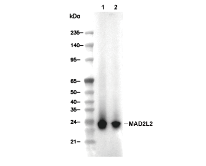 Lane 1: NCI-H1963, Lane 2: NCCIT
当該製品は品切れ状态で、メールアドレスをご教示いただければ、お客様に返信いたします。
代表番号: 045-509-1970|電子メール:sales@selleck.co.jp
キーポイント
WB
転写条件(ウェット): 200 mA, 60 min。
転写条件(ウェット): 200 mA, 60 min。
使用情報
| Dilution |
|---|
|
| Application |
|---|
| WB, IP |
| Source |
|---|
| Rabbit Monoclonal Antibody |
| Reactivity |
|---|
| Human, Mouse, Rat |
| Storage Buffer |
|---|
| PBS, pH 7.2+50% Glycerol+0.05% BSA+0.01% NaN3 |
| Storage (from the date of receipt) |
|---|
| -20°C (avoid freeze-thaw cycles), 2 years |
| Predicted MW |
|---|
| 24 kDa |
| ポジティブコントロール | NCI-H1963 cells; NCCIT cells; Calu cells; NMuMG cells; MBT2 cells; MTLn3 cells |
|---|---|
| ネガティブコントロール |
プロトコール
| WB |
|---|
Experimental Protocol:
Sample preparation
1. Tissue: Lyse the tissue sample by adding an appropriate volume of ice-cold RIPA/NP-40 Lysis Buffer (containing Protease Inhibitor Cocktail),and homogenize the tissue at a low temperature or lyse it by sonication on ice, then incubate on ice for 30 minutes. 2. Adherent cell: Aspirate the culture medium and wash the cells with ice-cold PBS twice. Lyse the cells by adding an appropriate volume of RIPA/NP-40 Lysis Buffer (containing Protease Inhibitor Cocktail), sonicate to lyse the cells, and incubate on ice for 30 minutes. 3. Suspension cell: Transfer the culture medium to a pre-cooled centrifuge tube. Centrifuge and aspirate the supernatant. Wash the cells with ice-cold PBS twice. Lyse the cells by adding an appropriate volume of RIPA/NP-40 Lysis Buffer (containing Protease Inhibitor Cocktail), sonicate to lyse the cells, and incubate on ice for 30 minutes. 4. Place the lysate into a pre-cooled microcentrifuge tube. Centrifuge at 4°C for 15 min. Collect the supernatant;
5. Remove a small volume of lysate to determine the protein concentration;
6. Combine the lysate with protein loading buffer. Boil 20 µL sample under 95-100°C for 5 min. Centrifuge for 5 min after cool down on ice.
Electrophoretic separation
1. According to the concentration of extracted protein, load appropriate amount of protein sample and marker onto SDS-PAGE gels for electrophoresis. Recommended separating gel (lower gel) concentration: 10%. Reference Table for Selecting SDS-PAGE Separation Gel Concentrations 2. Power up 80V for 30 minutes. Then the power supply is adjusted (110 V~150 V), the Marker is observed, and the electrophoresis can be stopped when the indicator band of the predyed protein Marker where the protein is located is properly separated. (Note that the current should not be too large when electrophoresis, too large current (more than 150 mA) will cause the temperature to rise, affecting the result of running glue. If high currents cannot be avoided, an ice bath can be used to cool the bath.)
Transfer membrane
1. Take out the converter, soak the clip and consumables in the pre-cooled converter;
2. Activate PVDF membrane with methanol for 1 min and rinse with transfer buffer;
3. Install it in the order of "black edge of clip - sponge - filter paper - filter paper - glue -PVDF membrane - filter paper - filter paper - sponge - white edge of clip"; 4. The protein was electrotransferred to PVDF membrane. ( 0.45 µm PVDF membrane is recommended ) Reference Table for Selecting PVDF Membrane Pore Size Specifications Recommended conditions for wet transfer: 200 mA, 60 min. ( Note that the transfer conditions can be adjusted according to the protein size. For high-molecular-weight proteins, a higher current and longer transfer time are recommended. However, ensure that the transfer tank remains at a low temperature to prevent gel melting.)
Block
1. After electrotransfer, wash the film with TBST at room temperature for 5 minutes;
2. Incubate the film in the blocking solution for 1 hour at room temperature;
3. Wash the film with TBST for 3 times, 5 minutes each time.
Antibody incubation
1. Use 5% skim milk powder to prepare the primary antibody working liquid (recommended dilution ratio for primary antibody 1:1000), gently shake and incubate with the film at 4°C overnight; 2. Wash the film with TBST 3 times, 5 minutes each time;
3. Add the secondary antibody to the blocking solution and incubate with the film gently at room temperature for 1 hour;
4. After incubation, wash the film with TBST 3 times for 5 minutes each time.
Antibody staining
1. Add the prepared ECL luminescent substrate (or select other color developing substrate according to the second antibody) and mix evenly;
2. Incubate with the film for 1 minute, remove excess substrate (keep the film moist), wrap with plastic film, and expose in the imaging system. |
生物学的記述
| Specificity |
|---|
| MAD2L2 Antibody (Rabbit mAb) [P22A23] detects endogenous levels of total MAD2L2 protein. |
| タンパク質の局在 |
|---|
| 染色体、細胞質、細胞骨格、細胞核 |
| Uniprot ID |
|---|
| Q9UI95 |
| Clone |
|---|
| P22A23 |
| Synonym(s) |
|---|
| Mitotic spindle assembly checkpoint protein MAD2B; MAD2-like protein 2; REV7; MAD2L2 |
| Background |
|---|
| Mitotic arrest deficient 2 like 2 (MAD2L2, also known as REV7) is a multifunctional HORMA‑domain protein that integrates cell‑cycle control with DNA damage response by participating in the spindle checkpoint machinery and several DNA repair pathways. It belongs to the MAD2‑like or HORMA family and adopts a closing‑clamshell architecture that enables it to act as an adaptor or scaffold, engaging partner proteins through interface‑switching modes rather than forming the core of the spindle assembly checkpoint itself. MAD2L2 functions as a core subunit of DNA polymerase ζ, where it bridges the catalytic subunit REV3 and the TLS scaffold REV1, coordinating lesion bypass during translesion synthesis and enabling replication through blocking DNA adducts. In double‑strand break repair, MAD2L2 joins REV1 and polymerase ζ at stalled forks and contributes to repair‑synthesis steps, while also participating in the shieldin complex, where it helps regulate pathway choice by limiting 5ʹ end resection and thereby favoring non‑homologous end joining over homologous recombination. MAD2L2 also contributes to mitotic regulation by restraining premature activation of the anaphase‑promoting complex/cyclosome through interactions with CDH1 and related checkpoint proteins, thereby helping to delay mitotic progression and maintain genome integrity under replication stress. MAD2L2 is frequently overexpressed compared with adjacent normal tissue, and high message or protein levels correlate with increased numbers of aberrant mitotic figures, chromosomal instability, and reduced overall survival in colorectal cancer and other solid malignancies, suggesting that elevated MAD2L2 expression reinforces error‑prone DNA repair and checkpoint adaptations that support tumor evolution. |
| References |
|---|
技術サポート
ストックの作り方、阻害剤の保管方法、細胞実験や動物実験の際に注意すべき点など、製品を取扱う時に問い合わせが多かった質問に対しては取扱説明書でお答えしています。
他に質問がある場合は、お気軽にお問い合わせください。
* 必須
納期 国内在庫品:受注日の翌日(15時までの受注分) *北海道、九州、沖縄への配送は受注日より2日以上 を要する場合あり 海外在庫品:受注後1〜2週間