
- 阻害剤
- 研究分野別
- PI3K/Akt/mTOR
- Epigenetics
- Methylation
- Immunology & Inflammation
- Protein Tyrosine Kinase
- Angiogenesis
- Apoptosis
- Autophagy
- ER stress & UPR
- JAK/STAT
- MAPK
- Cytoskeletal Signaling
- Cell Cycle
- TGF-beta/Smad
- 化合物ライブラリー
- 抗体
- 新製品
- お問い合わせ
Nav1.8/SCN10A Antibody [F24B8]
Catalog No.: F3462
Application:
Reactivity:
-
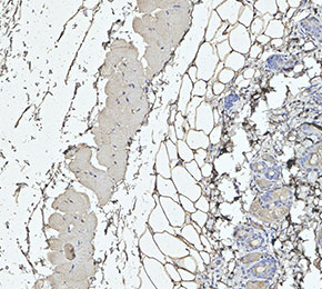 Immunohistochemical analysis of formalin fixed paraffin embedded mouse skin tissue with F3462 at 1:100 dilution.
使用情報
| Dilution |
|---|
|
| Application |
|---|
| IHC |
| Source |
|---|
| Mouse Monoclonal Antibody |
| Reactivity |
|---|
| Mouse, Rat |
| Storage Buffer |
|---|
| PBS, pH 7.2+50% Glycerol+0.05% BSA+0.01% NaN3 |
| Storage (from the date of receipt) |
|---|
| -20°C (avoid freeze-thaw cycles), 2 years |
| ポジティブコントロール | Mouse dorsal root ganglia; Rat dorsal root ganglia |
|---|---|
| ネガティブコントロール |
プロトコール
| IHC |
|---|
Experimental Protocol:
Deparaffinization/Rehydration
1. Deparaffinize/hydrate sections:
2. Incubate sections in three washes of xylene for 5 min each.
3. Incubate sections in two washes of 100% ethanol for 10 min each.
4. Incubate sections in two washes of 95% ethanol for 10 min each.
5. Wash sections two times in dH2O for 5 min each.
6.Antigen retrieval: For Citrate: Heat slides in a microwave submersed in 1X citrate unmasking solution until boiling is initiated; continue with 10 min at a sub-boiling temperature (95°-98°C). Cool slides on bench top for 30 min.
Staining
1. Wash sections in dH2O three times for 5 min each.
2. Incubate sections in 3% hydrogen peroxide for 10 min.
3. Wash sections in dH2O two times for 5 min each.
4. Wash sections in wash buffer for 5 min.
5. Block each section with 100–400 µl of blocking solution for 1 hr at room temperature.
6. Remove blocking solution and add 100–400 µl primary antibody diluent in to each section. Incubate overnight at 4°C.
7. Remove antibody solution and wash sections with wash buffer three times for 5 min each.
8. Cover section with 1–3 drops HRPas needed. Incubate in a humidified chamber for 30 min at room temperature.
9. Wash sections three times with wash buffer for 5 min each.
10. Add DAB Chromogen Concentrate to DAB Diluent and mix well before use.
11. Apply 100–400 µl DAB to each section and monitor closely. 1–10 min generally provides an acceptable staining intensity.
12. Immerse slides in dH2O.
13. If desired, counterstain sections with hematoxylin.
14. Wash sections in dH2O two times for 5 min each.
15. Dehydrate sections: Incubate sections in 95% ethanol two times for 10 sec each; Repeat in 100% ethanol, incubating sections two times for 10 sec each; Repeat in xylene, incubating sections two times for 10 sec each.
16. Mount sections with coverslips and mounting medium.
|
生物学的記述
| Specificity |
|---|
Nav1.8/SCN10A Antibody [F24B8] detects endogenous levels of total Nav1.8/SCN10A protein. |
| タンパク質の局在 |
|---|
| 細胞膜、細胞内膜系 |
| Uniprot ID |
|---|
| Q9Y5Y9 |
| Clone |
|---|
| F24B8 |
| Synonym(s) |
|---|
| Sodium channel protein type 10 subunit alpha, Peripheral nerve sodium channel 3, Sodium channel protein type X subunit alpha, Voltage-gated sodium channel subunit alpha Nav1.8, PN3, hPN3, SCN10A |
| Background |
|---|
| Nav1.8/SCN10A, encoded by the SCN10A gene, is a voltage-gated sodium channel α-subunit belonging to the TTX-resistant class, characterized by a cysteine substitution in its pore region that confers resistance to nanomolar tetrodotoxin inhibition. Structurally, it is a large transmembrane protein with four homologous domains (I–IV), each containing six membrane-spanning segments (S1–S6) that form the voltage-sensing and pore-forming regions, and is often associated with β-subunits that modulate gating and trafficking. NaV1.8 is predominantly expressed in small- and medium-diameter nociceptive sensory neurons of dorsal root ganglia (DRG) and cranial sensory ganglia, but is also present in peripheral neural elements of the heart, particularly intracardiac ganglia, while absent from cardiomyocytes. Functionally, NaV1.8 activates at relatively depolarized potentials, inactivates slowly, and maintains sodium currents at subthreshold voltages, enabling repetitive action potential firing and sustained excitability, especially at low temperatures. In DRG neurons, it plays a key role in pain signaling by amplifying excitability near the action potential threshold and interacting with NaV1.7, whereas in intracardiac neurons it regulates firing frequency and neurotransmitter release, potentially modulating cardiac conduction through neural control rather than direct myocyte depolarization. |
| References |
|---|
技術サポート
ストックの作り方、阻害剤の保管方法、細胞実験や動物実験の際に注意すべき点など、製品を取扱う時に問い合わせが多かった質問に対しては取扱説明書でお答えしています。
他に質問がある場合は、お気軽にお問い合わせください。
* 必須
納期 国内在庫品:受注日の翌日(15時までの受注分) *北海道、九州、沖縄への配送は受注日より2日以上 を要する場合あり 海外在庫品:受注後1〜2週間