
- 阻害剤
- 研究分野別
- PI3K/Akt/mTOR
- Epigenetics
- Methylation
- Immunology & Inflammation
- Protein Tyrosine Kinase
- Angiogenesis
- Apoptosis
- Autophagy
- ER stress & UPR
- JAK/STAT
- MAPK
- Cytoskeletal Signaling
- Cell Cycle
- TGF-beta/Smad
- 化合物ライブラリー
- 抗体
- 新製品
- お問い合わせ
NPRL2 Antibody [L19L7]
Catalog No.: F8198
Application:
Reactivity:
-
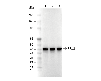 Lane 1: LnCAP, Lane 2: DLD-1, Lane 3: MCF7
使用情報
| Dilution |
|---|
|
| Application |
|---|
| WB, IP |
| Source |
|---|
| Rabbit Monoclonal Antibody |
| Reactivity |
|---|
| Human, Mouse, Rat, Monkey |
| Storage Buffer |
|---|
| PBS, pH 7.2+50% Glycerol+0.05% BSA+0.01% NaN3 |
| Storage (from the date of receipt) |
|---|
| -20°C (avoid freeze-thaw cycles), 2 years |
| Predicted MW |
|---|
| 41 kDa |
| ポジティブコントロール | LNCaP cells; DLD-1 cells; MCF-7 cells; IGROV-1 cells |
|---|---|
| ネガティブコントロール |
プロトコール
| WB |
|---|
Experimental Protocol:
Sample preparation
1. Tissue: Lyse the tissue sample by adding an appropriate volume of ice-cold RIPA/NP-40 Lysis Buffer (containing Protease Inhibitor Cocktail),and homogenize the tissue at a low temperature or lyse it by sonication on ice, then incubate on ice for 30 minutes. 2. Adherent cell: Aspirate the culture medium and wash the cells with ice-cold PBS twice. Lyse the cells by adding an appropriate volume of RIPA/NP-40 Lysis Buffer (containing Protease Inhibitor Cocktail), sonicate to lyse the cells, and incubate on ice for 30 minutes. 3. Suspension cell: Transfer the culture medium to a pre-cooled centrifuge tube. Centrifuge and aspirate the supernatant. Wash the cells with ice-cold PBS twice. Lyse the cells by adding an appropriate volume of RIPA/NP-40 Lysis Buffer (containing Protease Inhibitor Cocktail), sonicate to lyse the cells, and incubate on ice for 30 minutes. 4. Place the lysate into a pre-cooled microcentrifuge tube. Centrifuge at 4°C for 15 min. Collect the supernatant;
5. Remove a small volume of lysate to determine the protein concentration;
6. Combine the lysate with protein loading buffer. Boil 20 µL sample under 95-100°C for 5 min. Centrifuge for 5 min after cool down on ice.
Electrophoretic separation
1. According to the concentration of extracted protein, load appropriate amount of protein sample and marker onto SDS-PAGE gels for electrophoresis. Recommended separating gel (lower gel) concentration: 10%. Reference Table for Selecting SDS-PAGE Separation Gel Concentrations 2. Power up 80V for 30 minutes. Then the power supply is adjusted (110 V~150 V), the Marker is observed, and the electrophoresis can be stopped when the indicator band of the predyed protein Marker where the protein is located is properly separated. (Note that the current should not be too large when electrophoresis, too large current (more than 150 mA) will cause the temperature to rise, affecting the result of running glue. If high currents cannot be avoided, an ice bath can be used to cool the bath.)
Transfer membrane
1. Take out the converter, soak the clip and consumables in the pre-cooled converter;
2. Activate PVDF membrane with methanol for 1 min and rinse with transfer buffer;
3. Install it in the order of "black edge of clip - sponge - filter paper - filter paper - glue -PVDF membrane - filter paper - filter paper - sponge - white edge of clip"; 4. The protein was electrotransferred to PVDF membrane. ( 0.45 µm PVDF membrane is recommended ) Reference Table for Selecting PVDF Membrane Pore Size Specifications Recommended conditions for wet transfer: 200 mA, 120 min. ( Note that the transfer conditions can be adjusted according to the protein size. For high-molecular-weight proteins, a higher current and longer transfer time are recommended. However, ensure that the transfer tank remains at a low temperature to prevent gel melting.)
Block
1. After electrotransfer, wash the film with TBST at room temperature for 5 minutes;
2. Incubate the film in the blocking solution for 1 hour at room temperature;
3. Wash the film with TBST for 3 times, 5 minutes each time.
Antibody incubation
1. Use 5% skim milk powder to prepare the primary antibody working liquid (recommended dilution ratio for primary antibody 1:1000), gently shake and incubate with the film at 4°C overnight; 2. Wash the film with TBST 3 times, 5 minutes each time;
3. Add the secondary antibody to the blocking solution and incubate with the film gently at room temperature for 1 hour;
4. After incubation, wash the film with TBST 3 times for 5 minutes each time.
Antibody staining
1. Add the prepared ECL luminescent substrate (or select other color developing substrate according to the second antibody) and mix evenly;
2. Incubate with the film for 1 minute, remove excess substrate (keep the film moist), wrap with plastic film, and expose in the imaging system. |
生物学的記述
| Specificity |
|---|
| NPRL2 Antibody [L19L7] detects endogenous levels of total NPRL2 protein. |
| タンパク質の局在 |
|---|
| リソソーム、細胞内膜系 |
| Uniprot ID |
|---|
| Q8WTW4 |
| Clone |
|---|
| L19L7 |
| Synonym(s) |
|---|
| GATOR1 complex protein NPRL2; Gene 21 protein 1 publication (G21 protein); Nitrogen permease regulator 2-like protein (NPR2-like protein); Tumor suppressor candidate 4; NPRL2; TUSC4 |
| Background |
|---|
| NPRL2 (also known as TUSC4) is a core component of the GATOR1 complex (with DEPDC5 and NPRL3), functioning as a negative regulator of mTORC1 signaling by acting as a GAP (GTPase-activating protein) for RagA/B GTPases, thereby inhibiting amino acid-induced mTORC1 translocation to lysosomes under nutrient limitation to suppress anabolic processes like protein synthesis. NPRL2 features an N-terminal Longin domain (residues ~1–180) for Rag GTPase binding and GAP activity, a central helical domain facilitating DEPDC5/NPRL3 heterodimerization, and a C-terminal region enabling lysosomal membrane association and selective Raptor interaction; key residues in the Longin domain (e.g., conserved Arg/Lys motifs, notably Arg78) catalyze GTP hydrolysis on RagA GDP/RagD GTP heterodimers. During amino acid sufficiency NPRL2 paradoxically interacts with Raptor to fine-tune mTORC1 activation via a "seesaw" mechanism, releasing Rag inhibition while preventing overactivation, while amino acid scarcity shifts NPRL2-Rag binding to robustly suppress mTORC1, maintaining metabolic homeostasis. NPRL2 overexpression inhibits cell proliferation by sustaining BRCA1 stability and inducing ROS via NOX2/mitochondrial dysfunction, triggering DNA damage responses and apoptosis in p53-proficient cells. NPRL2 ensures neurometabolic balance (e.g., regulating sodium channel expression in neurons), prevents excessive growth under stress, and acts as a tumor suppressor limiting mTORC1-driven oncogenesis. Pathogenic germline mutations cause familial focal epilepsies and mTORopathies (e.g., cortical dysplasia) due to hyperactive mTORC1, while somatic loss promotes tumorigenesis. |
| References |
|---|
技術サポート
ストックの作り方、阻害剤の保管方法、細胞実験や動物実験の際に注意すべき点など、製品を取扱う時に問い合わせが多かった質問に対しては取扱説明書でお答えしています。
他に質問がある場合は、お気軽にお問い合わせください。
* 必須
納期 国内在庫品:受注日の翌日(15時までの受注分) *北海道、九州、沖縄への配送は受注日より2日以上 を要する場合あり 海外在庫品:受注後1〜2週間