
- 阻害剤
- 研究分野別
- PI3K/Akt/mTOR
- Epigenetics
- Methylation
- Immunology & Inflammation
- Protein Tyrosine Kinase
- Angiogenesis
- Apoptosis
- Autophagy
- ER stress & UPR
- JAK/STAT
- MAPK
- Cytoskeletal Signaling
- Cell Cycle
- TGF-beta/Smad
- 化合物ライブラリー
- 抗体
- 新製品
- お問い合わせ
Phospho-Hsp27 (Ser82) Antibody [M1P19]
Catalog No.: F3764
Application:
Reactivity:
-
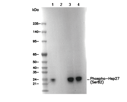 Lane 1: HeLa (with 44°C heat shock), Lane 2: HeLa, Lane 3: Mouse heart, Lane 4: Rat heart
キーポイント
WB
転写条件(ウェット): 200 mA, 60 min。
転写条件(ウェット): 200 mA, 60 min。
使用情報
| Dilution |
|---|
|
| Application |
|---|
| WB, IP |
| Source |
|---|
| Rabbit Monoclonal Antibody |
| Reactivity |
|---|
| Mouse, Rat, Human |
| Storage Buffer |
|---|
| PBS, pH 7.2+50% Glycerol+0.05% BSA+0.01% NaN3 |
| Storage (from the date of receipt) |
|---|
| -20°C (avoid freeze-thaw cycles), 2 years |
| Predicted MW Observed MW |
|---|
| 23 kDa 27 kDa |
| *なぜ予測分子量と実際の分子量が異なるのか? 下記の原因により、実際の分子量が予測と異なる:タンパク質の翻訳後修飾(リン酸化/糖鎖付加),スプライシングバリアント,イソフォーム,相対的な電荷,ポリマー。 |
| ポジティブコントロール | Mouse heart; Rat heart; HeLa (with 44°C heat shock treatment) |
|---|---|
| ネガティブコントロール |
プロトコール
| WB |
|---|
Experimental Protocol:
Sample preparation
1. Tissue: Lyse the tissue sample by adding an appropriate volume of ice-cold RIPA/NP-40 Lysis Buffer (containing Protease Inhibitor Cocktail, Phosphatase Inhibitor Cocktail),and homogenize the tissue at a low temperature or lyse it by sonication on ice, then incubate on ice for 30 minutes. 2. Adherent cell: Aspirate the culture medium and wash the cells with ice-cold PBS twice. Lyse the cells by adding an appropriate volume of RIPA/NP-40 Lysis Buffer (containing Protease Inhibitor Cocktail, Phosphatase Inhibitor Cocktail), sonicate to lyse the cells, and incubate on ice for 30 minutes. 3. Suspension cell: Transfer the culture medium to a pre-cooled centrifuge tube. Centrifuge and aspirate the supernatant. Wash the cells with ice-cold PBS twice. Lyse the cells by adding an appropriate volume of RIPA/NP-40 Lysis Buffer (containing Protease Inhibitor Cocktail, Phosphatase Inhibitor Cocktail), sonicate to lyse the cells, and incubate on ice for 30 minutes. 4. Place the lysate into a pre-cooled microcentrifuge tube. Centrifuge at 4°C for 15 min. Collect the supernatant;
5. Remove a small volume of lysate to determine the protein concentration;
6. Combine the lysate with protein loading buffer. Boil 20 µL sample under 95-100°C for 5 min. Centrifuge for 5 min after cool down on ice.
Electrophoretic separation
1. According to the concentration of extracted protein, load appropriate amount of protein sample and marker onto SDS-PAGE gels for electrophoresis. Recommended separating gel (lower gel) concentration: 10%. Reference Table for Selecting SDS-PAGE Separation Gel Concentrations 2. Power up 80V for 30 minutes. Then the power supply is adjusted (110 V~150 V), the Marker is observed, and the electrophoresis can be stopped when the indicator band of the predyed protein Marker where the protein is located is properly separated. (Note that the current should not be too large when electrophoresis, too large current (more than 150 mA) will cause the temperature to rise, affecting the result of running glue. If high currents cannot be avoided, an ice bath can be used to cool the bath.)
Transfer membrane
1. Take out the converter, soak the clip and consumables in the pre-cooled converter;
2. Activate PVDF membrane with methanol for 1 min and rinse with transfer buffer;
3. Install it in the order of "black edge of clip - sponge - filter paper - filter paper - glue -PVDF membrane - filter paper - filter paper - sponge - white edge of clip"; 4. The protein was electrotransferred to PVDF membrane. ( 0.45 µm PVDF membrane is recommended ) Reference Table for Selecting PVDF Membrane Pore Size Specifications Recommended conditions for wet transfer: 200 mA, 60 min. ( Note that the transfer conditions can be adjusted according to the protein size. For high-molecular-weight proteins, a higher current and longer transfer time are recommended. However, ensure that the transfer tank remains at a low temperature to prevent gel melting.)
Block
1. After electrotransfer, wash the film with TBST at room temperature for 5 minutes;
2. Incubate the film in the blocking solution ( recommending 5% BSA solution)
for 1 hour at room temperature;
3. Wash the film with TBST for 3 times, 5 minutes each time.
Antibody incubation
1. Use 5% skim milk powder to prepare the primary antibody working liquid (recommended dilution ratio for primary antibody 1:1000), gently shake and incubate with the film at 4°C overnight; 2. Wash the film with TBST 3 times, 5 minutes each time;
3. Add the secondary antibody to the blocking solution and incubate with the film gently at room temperature for 1 hour;
4. After incubation, wash the film with TBST 3 times for 5 minutes each time.
Antibody staining
1. Add the prepared ECL luminescent substrate (or select other color developing substrate according to the second antibody) and mix evenly;
2. Incubate with the film for 1 minute, remove excess substrate (keep the film moist), wrap with plastic film, and expose in the imaging system. |
生物学的記述
| Specificity |
|---|
Phospho-Hsp27 (Ser82) Antibody [M1P19] detects endogenous levels of total Phospho-HSP27 (Ser82) protein only when phosphorylated at Ser82. |
| タンパク質の局在 |
|---|
| 細胞質、細胞骨格、細胞核 |
| Uniprot ID |
|---|
| P04792 |
| Clone |
|---|
| M1P19 |
| Synonym(s) |
|---|
| HSP27, HSP28, HSPB1, Heat shock protein beta-1, HspB1, 28 kDa heat shock protein, Estrogen-regulated 24 kDa protein, Heat shock 27 kDa protein, Heat shock protein family B member 1, Stress-responsive protein 27, HSP 27, SRP27 |
| Background |
|---|
Heat shock protein 27 (HSP27) is a member of the small heat shock protein (sHSP) family (12–43 kDa) and exhibits a wide range of cellular functions. It acts as a molecular chaperone, an antioxidant, a regulator of apoptosis, and a mediator of actin cytoskeletal remodeling. Like other small HSPs, HSP27 contains a conserved α-crystallin domain located within its C-terminal region. Under oxidative stress, HSP27 protects cells by lowering intracellular levels of reactive oxygen species (ROS), achieved through enhancing glutathione production and reducing free iron availability. It also exhibits strong anti-apoptotic activity, influencing both mitochondria-dependent and independent apoptotic pathways. For example, HSP27 binds to DAXX during Fas–FasL–mediated apoptosis, preventing DAXX from associating with ASK1. Additionally, it interacts with Bax and cytochrome c, thereby suppressing mitochondrial apoptosis and blocking caspase-dependent cell death. HSP27 is expressed at basal levels in most cells and tissues, where it typically exists as large oligomeric complexes. Upon exposure to stress, its expression rises significantly, enhancing cellular resistance to damaging conditions. The protein is subject to stress-induced phosphorylation at Ser15, Ser78, and Ser82 in humans (Ser15 and Ser86 in rodents), mediated by MAPKAP kinase 2/3 downstream of the p38 MAPK signaling pathway. Phosphorylation not only influences its oligomeric state but also regulates its interactions with partner proteins. In neutrophils, HSP27 forms a complex with AKT and MAPKAP kinase 2, which suppresses constitutive apoptosis and promotes an inflammatory response. Stress-induced phosphorylation of HSP27 disrupts this complex, leading to dissociation from AKT and restoration of neutrophil apoptosis. Notably, the effects of phosphorylated HSP27 are highly context-dependent, varying with cell type and signaling environment. |
| References |
|---|
技術サポート
ストックの作り方、阻害剤の保管方法、細胞実験や動物実験の際に注意すべき点など、製品を取扱う時に問い合わせが多かった質問に対しては取扱説明書でお答えしています。
他に質問がある場合は、お気軽にお問い合わせください。
* 必須
納期 国内在庫品:受注日の翌日(15時までの受注分) *北海道、九州、沖縄への配送は受注日より2日以上 を要する場合あり 海外在庫品:受注後1〜2週間